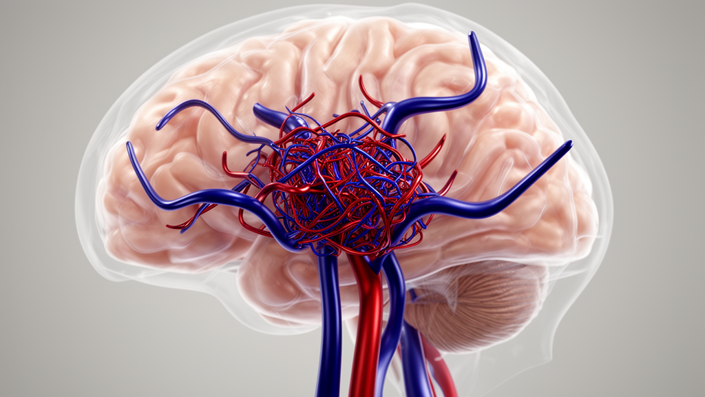
Brain arteriovenous malformations, tangles of dysplastic vessels in which arteries connect directly to veins without an intervening capillary network, are usually described by their feeding arteries or their nidus. Far less attention has been paid to the veins that carry blood away from them, even though these draining veins can be the source of hemorrhage and can be the target of specific treatments. A new anatomical study, published in the Egyptian Journal of Neurosurgery, now provides one of the most detailed radiologic portraits of these vessels to date, based on 57 patients examined with digital subtraction angiography and computed tomographic angiography.
The research team, led by Biljana Milatović and Svetlana Valjarević with colleagues including Slobodan Marinković, enrolled patients at the Neuroradiologic Department of a Neurosurgical Clinic between January 2019 and February 2021. Every participant received a neurological examination and routine biochemical analyses before imaging. The group averaged 35.46 years of age, with a standard deviation of 15.64 years, and mainly ranged from 20 to 45 years old. The female-to-male ratio was almost equal. More than one third of the patients had hypertension, at 35.1 percent, and 17.6 percent had diabetes mellitus, although neither association reached statistical significance in the analysis.
The imaging protocol was thorough. Digital subtraction angiography was performed by transfemoral catheterisation with automatic bolus injection of contrast agent in volumes of 5 to 8 milliliters per sequence, producing a serial four-vessel cerebral angiogram on a monoplane Siemens Axiom Artis unit with rotational capabilities. Three-dimensional reconstructions were generated from up to 12 milliliters of contrast injected at 2.5 milliliters per second. Computed tomographic angiography used a 128-slice Siemens Somatom Definition AS scanner with 100 milliliters of Ultravist 370 contrast delivered at 4.5 milliliters per second, followed by volume rendering and multipolar reconstruction of the vessels and skull. Every image set was read independently by two neuroradiologists and one neuroanatomist, and the data were analyzed statistically with SPSS software.
The malformations themselves showed a clear anatomical pattern. The nidus, the convolution of tortuous vessels at the heart of the malformation, was most often located in the frontal region, in 17.6 percent of patients, with the temporal and parietotemporal areas each accounting for 15.8 percent. A superficial, pial position on the brain surface was the most common, seen in 63.2 percent of cases, compared with a deep location in 12.3 percent and a combination of the two in 24.5 percent. Supratentorial malformations, those above the tentorium of the cerebellum, dominated at 88.4 percent, while 12.6 percent lay infratentorially. The nidus averaged 21.5 by 28.5 millimeters, a finding the authors report as statistically significant.
The central finding of the study concerns the draining veins themselves. Across the 57 patients, the number of draining veins ranged from 1 to 17, with a mean of 2.19, a distribution the authors report as statistically significant. A single draining vein was present in 22.8 percent of patients, two veins in 59.7 percent, and multiple veins in the remaining 17.6 percent. Where the veins terminated mattered as much as how many there were. Most, 63.2 percent, emptied into the superior sagittal sinus and other superficial vessels, while 12.3 percent terminated in deep veins or sinuses and 24.5 percent drained into both superficial and deep systems.
Among the individual recipient vessels, the superior sagittal sinus stood out as the most frequent single drainage site, receiving venous blood alone in 38.6 percent of patients. The transverse sinus and the straight sinus each served as the sole drainage route in 7.2 percent, and the sigmoid sinus in 3.6 percent. The sphenoparietal sinus, the cavernous sinus, the petrosal sinuses, and the confluence of sinuses each appeared as the sole drainage site in a single case, or 1.8 percent of the cohort. The authors note that draining veins may be aberrant vessels or modified versions of standard veins, including pial anastomoses such as the veins of Trolard and Labbé, and occasionally deep venous connections that link superficial veins with white matter vessels and onward to the striate or thalamic veins.
The morphology of these vessels was striking. Among the 128 draining veins registered in the study, tortuous vessels were by far the most common shape, accounting for 45.4 percent, while straight, angular, arch-shaped or S-shaped, spiral, and bifurcated forms appeared less often, some with loops. Dilation, or ectasia, was nearly universal: 88.9 percent of patients had dilated draining veins. Venous aneurysms, which the authors describe as singular or multiple in affected patients, were present in 8.8 percent. One vein in the series was exceptionally long, coursing from near the frontal pole all the way to the ipsilateral transverse sinus. In the discussion, the authors report that draining veins averaged 11 millimeters in diameter, with a standard deviation of 5.3 millimeters, a size commonly associated with higher blood flow volume and pressure.
Hemorrhage emerged as a frequent first sign of the malformations. Bleeding, including venous bleeding, was the initial manifestation in 49.2 percent of patients, taking the form of intracerebral, intraventricular, or subarachnoid hemorrhage, or combinations of these. The statistical analysis linked bleeding patterns to vascular features: hemorrhage occurred in 22.8 percent of patients with nidus aneurysms, a statistically significant association, and in 54.4 percent of those with dilated draining veins. Bleeding was also more frequent in small malformations, at 45.7 percent, and repeated hemorrhage was rare, at 5.3 percent. These figures align with the broader literature the authors cite, which estimates a 2 to 4 percent annual risk of hemorrhage associated with these malformations, particularly affecting deep and infratentorial lesions, those with smaller nidus volume, those with only one or two draining veins, and those with associated aneurysms.
The pathophysiological explanation offered by the authors rests on hemodynamics. Because the malformations lack a regular capillary network, blood shunts at high flow directly from arteries to veins; the authors note that local cerebral blood flow within these lesions averages about 490 milliliters per 100 grams per minute, compared with a normal value of 55. This high, often turbulent flow promotes venous dilation and tortuosity, raises intravascular pressure and wall shear stress, and can produce local ischemia and a steal phenomenon in the surrounding brain. The vessel walls themselves may show endothelial dysfunction, lipid deposition, hyalinization, damage to the internal elastic lamina, hyperplasia of altered smooth muscle cells, fibrosis, and signs of inflammation, changes that can weaken the wall and lead to hemorrhage, and rarely to stenosis or occlusion. The authors also point out that enhanced vein wall visibility on imaging, together with reduced blood velocity and wall shear stress, could help predict draining vein stenosis, which they associate with a mean diameter of 2.9 millimeters and a consequent risk of hemorrhage from the nidus.
Treatment in the cohort reflected the range of options used in contemporary practice. Endovascular embolization was performed in 15.8 percent of patients, surgical intervention in 12.3 percent, and gamma knife irradiation in 21.1 percent, while the remaining patients were treated conservatively. Despite this mix, the total mortality rate in the group was 7.2 percent, which sits within the reported range of 5 to 22 percent for ruptured malformations cited by the authors. The study’s contribution is anatomical rather than therapeutic: by systematically documenting how many draining veins exist, where they terminate, and how often they dilate, tortuous or aneurysmal, it gives clinicians a detailed radiologic reference for a vascular compartment that is therapeutically and prognostically important, including for emerging transvenous interventions. The authors acknowledge that their single-center cohort of 57 patients, imaged with DSA and CTA over a two-year window, is a starting point for a topic they describe as scarcely covered in the available literature.
Subject of Research: The radiologic anatomy of the draining veins of cerebral arteriovenous malformations in 57 patients examined with digital subtraction angiography and CT angiography.
Article Title: Radiologic anatomy of draining veins of the cerebral arteriovenous malformations (AVMs)
Article References: Milatović, B., Valjarević, S., Milić, I., Damjanović, G., Djulejić, V., Georgievski-Brkić, B., Blagojević, V., & Marinković, S. (2026). Radiologic anatomy of draining veins of the cerebral arteriovenous malformations (AVMs). Egyptian Journal of Neurosurgery, 41(1), Article 175. https://doi.org/10.1186/s41984-026-00683-y
Image Credits: AI Generated
DOI: 10.1186/s41984-026-00683-y
Keywords: cerebral arteriovenous malformation, draining veins, digital subtraction angiography, CT angiography, venous aneurysm, intracranial hemorrhage, superior sagittal sinus, vascular anatomy, endovascular embolization, gamma knife radiosurgery, neuroradiology, venous ectasia
Cite Scienmag News
Cassandra Pierce. (October 28, 2026). Draining Veins of Brain AVMs Mapped in 57 Patients Reveal Dilated, Tortuous Vessels. Scienmag. https://scienmag.com/draining-veins-of-brain-avms-mapped-in-57-patients-reveal-dilated-tortuous-vessels/
Cassandra Pierce. "Draining Veins of Brain AVMs Mapped in 57 Patients Reveal Dilated, Tortuous Vessels." Scienmag, 28 October 2026, https://scienmag.com/draining-veins-of-brain-avms-mapped-in-57-patients-reveal-dilated-tortuous-vessels/. Accessed 29 October 2026.
Cassandra Pierce. "Draining Veins of Brain AVMs Mapped in 57 Patients Reveal Dilated, Tortuous Vessels." Scienmag. October 28, 2026. https://scienmag.com/draining-veins-of-brain-avms-mapped-in-57-patients-reveal-dilated-tortuous-vessels/