Friday, October 30, 2026
Science
No Result
View All Result
  • Login
  • HOME
  • SCIENCE NEWS
  • CONTACT US
  • HOME
  • SCIENCE NEWS
  • CONTACT US
No Result
View All Result
Scienmag
No Result
View All Result
Home Science News Biology

Microscopy Reveals Midgut and Malpighian Tubule Structure of Beetle Oxythyrea funesta

October 28, 2026
in Biology
Drew Townsend
By Drew Townsend Scienmag Editorial Profile - Cell Biology
Reading Time: 3 mins read
0
Microscopy Reveals Midgut and Malpighian Tubule Structure of Beetle Oxythyrea funesta

Microscopy Reveals Midgut and Malpighian Tubule Structure of Beetle Oxythyrea funesta

65
SHARES
587
VIEWS
Share on FacebookShare on Twitter
ADVERTISEMENT

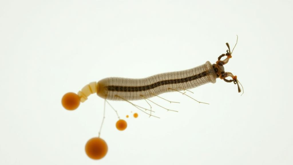
Researchers have mapped the fine structure of the digestive and excretory organs of the scarab beetle Oxythyrea funesta, using stereo, light, and scanning electron microscopy to document the midgut and Malpighian tubules in detail. The study, published in BMC Zoology, provides a morphological baseline for this species, which the authors say can support comparative work across the family Scarabaeidae and, ultimately, the development of selective pest management strategies.

In insects, the midgut is the primary organ where chemical digestion and nutrient absorption take place, while the Malpighian tubules remove metabolic waste and maintain osmoregulation.

The examination showed that the digestive tract of O. funesta is divided into three sections, with the midgut lying between the foregut and hindgut. The midgut proved to be the longest region, extending as a straight, tube-like structure from the foregut and forming two annular structures toward the hindgut.

Histologically, the midgut is characterized by a single-layered cylindrical epithelium, a thin muscle layer, and a peritrophic membrane within the lumen. The epithelial cells show apocrine secretion, and regenerative cells are present at the base of the epithelium. Under the scanning electron microscope, secretory vesicles could be seen in the lumen, along with short microvilli extending from the apical surface of the epithelial cells.

The Malpighian tubules, by contrast, are a pair of long, tubular structures with flat surfaces that run from the midgut to the hindgut. Light and scanning electron microscopy of these tubules revealed microvilli extending from the apical surface of a single-layered cuboidal epithelium whose cells carry round nuclei.

The authors state that the study aims to characterize the midgut and Malpighian tubules of O. funesta in a way that supports comparative studies within Scarabaeidae and future work on selective pest management. Because the article was shared early to provide faster access to peer-reviewed, accepted research, it is citable and carries a permanent DOI, though the version is subject to further edits before the final Version of Record.

Similar morphological and histological studies have been published on other beetles and insects, including work on the midgut and Malpighian tubules of Copris felschei, the alimentary canal and associated structures of Pyrrhocoris apterus, and the ultrastructure of midgut, Malpighian tubules, testis, and ovariole in the superworm Zophobas morio.

For O. funesta, the new description links gross anatomy, histology, and surface ultrastructure in a single account: a long, straight midgut with annular junctions, an apocrinally secreting cylindrical epithelium protected by a peritrophic membrane, and paired Malpighian tubules lined with cuboidal epithelium bearing apical microvilli. These features now provide a documented reference point for future anatomical and applied studies on this scarab species.

Subject of Research: Morphology of the midgut and Malpighian tubules of the scarab beetle Oxythyrea funesta examined by light and scanning electron microscopy

Article Title: A morphological study on the midgut and Malpighian tubules of Oxythyrea funesta (Poda, 1761) (Coleoptera: Scarabaeidae) using light and scanning electron microscopy

Article References: Çağlar, Ü., Arslan, H., Özyurt Koçakoğlu, N., & Candan, S. (2026). A morphological study on the midgut and Malpighian tubules of Oxythyrea funesta (Poda, 1761) (Coleoptera: Scarabaeidae) using light and scanning electron microscopy. BMC Zoology. https://doi.org/10.1186/s40850-026-00292-0

Image Credits: AI Generated

DOI: 10.1186/s40850-026-00292-0

Keywords: Oxythyrea funesta, Scarabaeidae, Coleoptera, midgut, Malpighian tubules, insect morphology, histology, scanning electron microscopy, peritrophic membrane, apocrine secretion, digestive system, excretory system

Cite Scienmag News

Drew Townsend. (October 28, 2026). Microscopy Reveals Midgut and Malpighian Tubule Structure of Beetle Oxythyrea funesta. Scienmag. https://scienmag.com/microscopy-reveals-midgut-and-malpighian-tubule-structure-of-beetle-oxythyrea-funesta/

Drew Townsend. "Microscopy Reveals Midgut and Malpighian Tubule Structure of Beetle Oxythyrea funesta." Scienmag, 28 October 2026, https://scienmag.com/microscopy-reveals-midgut-and-malpighian-tubule-structure-of-beetle-oxythyrea-funesta/. Accessed 30 October 2026.

Drew Townsend. "Microscopy Reveals Midgut and Malpighian Tubule Structure of Beetle Oxythyrea funesta." Scienmag. October 28, 2026. https://scienmag.com/microscopy-reveals-midgut-and-malpighian-tubule-structure-of-beetle-oxythyrea-funesta/

Tags: apocrine secretionbeetle digestive system microscopyColeopteracomparative beetle anatomydigestive systemexcretory systemhistologyhistology of insect midgutinsect excretory organs morphologyinsect morphologyinsect nutrient absorption anatomyinsect osmoregulation structuresinsect waste removal mechanismsMalpighian tubulesMalpighian tubules in scarab beetlesmidgutmorphological baseline for scarab beetlesOxythyrea funestaOxythyrea funesta midgut structureperitrophic membranepest management strategies in scarabaeidaescanning electron microscopyscanning electron microscopy of insect tissuesScarabaeidae
Share26Tweet16
Previous Post

Gut Microbiomes Diverge Between Wild and Semi-Captive Przewalski’s Gazelles Across Age Stages

Next Post

Rapid 40-minute fluorescent test detects optrA resistance gene in Enterococcaceae

Related Posts

Microbial Sources and Stress Shape the Hidden Endophytes of an Invasive Plant
Biology

Microbial Sources and Stress Shape the Hidden Endophytes of an Invasive Plant

October 30, 2026
Firebreaks Leave Lasting Scars in Grasslands While Wildfire Heals Fast
Biology

Firebreaks Leave Lasting Scars in Grasslands While Wildfire Heals Fast

October 30, 2026
Hidden Peptides in Non-Coding RNAs Reshape Cardiovascular Biology
Biology

Hidden Peptides in Non-Coding RNAs Reshape Cardiovascular Biology

October 30, 2026
Phage K Holin’s C-Terminal Region Shapes Antibacterial Activity, Study Finds
Biotechnology

Phage K Holin’s C-Terminal Region Shapes Antibacterial Activity, Study Finds

October 30, 2026
Computational screen flags five natural compounds that bind a tuberculosis virulence enzyme
Chemistry

Computational screen flags five natural compounds that bind a tuberculosis virulence enzyme

October 30, 2026
Antifungal liver injury study pinpoints KDR as shared hepatotoxicity target
Biology

Antifungal liver injury study pinpoints KDR as shared hepatotoxicity target

October 30, 2026
Next Post
Rapid 40-minute fluorescent test detects optrA resistance gene in Enterococcaceae

Rapid 40-minute fluorescent test detects optrA resistance gene in Enterococcaceae

  • Mothers who receive childcare support from maternal grandparents show more optimized

    Mothers who receive childcare support from maternal grandparents show more parental warmth, finds NTU Singapore study

    27656 shares
    Share 11059 Tweet 6912
  • University of Seville Breaks 120-Year-Old Mystery, Revises a Key Einstein Concept

    1061 shares
    Share 424 Tweet 265
  • Bee body mass, pathogens and local climate influence heat tolerance

    682 shares
    Share 273 Tweet 171
  • Researchers record first-ever images and data of a shark experiencing a boat strike

    546 shares
    Share 218 Tweet 137
  • Groundbreaking Clinical Trial Reveals Lubiprostone Enhances Kidney Function

    531 shares
    Share 212 Tweet 133
Science

Embark on a thrilling journey of discovery with Scienmag.com—your ultimate source for cutting-edge breakthroughs. Immerse yourself in a world where curiosity knows no limits and tomorrow’s possibilities become today’s reality!

RECENT NEWS

  • Microbial Sources and Stress Shape the Hidden Endophytes of an Invasive Plant
  • Firebreaks Leave Lasting Scars in Grasslands While Wildfire Heals Fast
  • Regularisation and voluntary return reproduce precarity for irregularised migrants in Spain and the UK
  • ZNF207 shields glutamine transporter ASCT2 from degradation to fuel liver cancer growth

Categories

  • Agriculture
  • Anthropology
  • Archaeology
  • Athmospheric
  • Biology
  • Biotechnology
  • Blog
  • Bussines
  • Cancer
  • Chemistry
  • Climate
  • Earth Science
  • Editorial Policy
  • Marine
  • Mathematics
  • Medicine
  • Pediatry
  • Policy
  • Psychology & Psychiatry
  • Science Education
  • Science News
  • Social Science
  • Space
  • Technology and Engineering

Subscribe to Blog via Email

Enter your email address to subscribe to this blog and receive notifications of new posts by email.

Join 5,150 other subscribers

© 2025 Scienmag - Science Magazine

Welcome Back!

Login to your account below

Forgotten Password?

Retrieve your password

Please enter your username or email address to reset your password.

Log In
No Result
View All Result
  • HOME
  • SCIENCE NEWS
  • CONTACT US

© 2025 Scienmag - Science Magazine

Discover more from Science

Subscribe now to keep reading and get access to the full archive.

Continue reading