The developmental gene engrailed has spent more than a century in the textbooks as a segmentation gene, famous for carving the body plans of fruit flies into repeating segments. A new study of the microscopic rotifer Brachionus manjavacas shows the gene doing something no one has ever documented before: switching on inside the excretory machinery of an animal barely visible to the naked eye. The finding, published in Discover Developmental Biology, adds a striking new entry to the long list of jobs this ancient transcription factor performs across the animal kingdom.
Andreas C. Fröbius, the study’s author, set out to fill a conspicuous gap. Engrailed has been studied intensively in arthropods, molluscs, brachiopods, echinoderms, annelids, onychophorans, tardigrades, cephalochordates, tunicates and vertebrates, yet nothing at all was known about its deployment in Gnathifera, a major spiralian clade that includes rotifers, gnathostomulids, micrognathozoans, acanthocephalans and chaetognaths. Rotifers are ideal for probing such questions because they combine extreme miniaturization with well-organized epithelial, ciliary and neurosecretory systems that can be mapped with cellular precision during embryonic development.
The first task was to confirm that the rotifer gene really is engrailed. Fröbius isolated a short homeodomain fragment from complementary DNA of mixed embryonic stages, then recovered the full-length transcript from a newly generated transcriptome of the same strain. The sequence has been deposited in GenBank under accession number PX666008, and the gene’s genomic position was verified against the Brachionus manjavacas RUS genome assembly. Phylogenetic analysis of 3,046 Engrailed proteins, built from the conserved engrailed homology regions EH1 through EH5, placed the gnathiferan sequences firmly within the protostome engrailed clade, confirming orthology. The tree comprised 2,020 deuterostome, 880 ecdysozoan, 109 lophotrochozoan and 37 gnathiferan sequences, and node support was assessed with 1,000 nonparametric bootstrap replicates.
The rotifer gene turned out to be structurally complete. The encoded protein spans 403 amino acids and carries the full canonical bilaterian motif complement: the EH1 repression motif that interacts with Groucho co-repressors, the EH2 motif that enables cooperative binding with TALE-class cofactors such as Pbx/Exd, the homeodomain itself, and the C-terminal EH5 signature. Both a Groucho ortholog and a predicted Pbx/Exd-like gene exist in the rotifer genome, so the interaction partners implied by the protein architecture are present. A single engrailed locus, as seen in Brachionus manjavacas, is likely the ancestral bilaterian condition, whereas arthropods carry a tandem engrailed–invected pair and vertebrates have the duplicated En1 and En2 genes.
One architectural detail stands out as an evolutionary landmark. An intron interrupts the coding sequence within the EH2 region at precisely the same position and phase in every examined protostome and deuterostome ortholog, indicating that this splice junction predates the protostome–deuterostome split. In Brachionus manjavacas the gene sits near the 5′ end of scaffold 8 and consists of two coding regions separated by a 581-base-pair intron. While the splice site itself is invariant, intron length varies enormously across taxa, from compact forms in some rotifers to very large introns in molluscs and echinoderms. The author suggests that intron position is evolutionarily constrained while intron size drifts freely with lineage-specific genomic architecture, a pattern that links engrailed gene structure across more than half a billion years of bilaterian evolution.
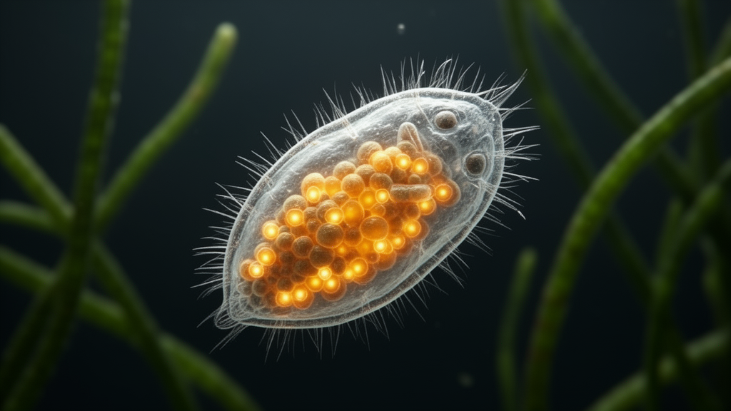
The expression data delivered the surprise. Using whole-mount in situ hybridization on amictic embryos, Fröbius found that at Stage 4 of development engrailed is active in a posterior domain that becomes restricted to the developing foot, marking two cells in its mid-region that coincide with the position of two known foot neurons. In addition, ventrolateral expression domains appear in the trunk region, shift slightly anteriorly, elongate and subdivide. By Stage 5 these domains have resolved into chains of individual connected cells spanning the posterior two-thirds of the trunk, and shortly before hatching four elongated rod-like domains are present on each side. Immunofluorescence staining of amictic females with an anti-Engrailed/Invected antibody confirmed the pattern and revealed the protein’s precise location: the terminal organs, the flame cells of the protonephridial system, the nuclei of the canal cells, and Huxley’s anastomose, a transverse anterior renal commissure connecting the two protonephridia laterally and dorsally. Expression in the foot is downregulated after hatching, while the protonephridial signal persists in adult females.
That localization is genuinely novel. Engrailed expression in excretory or protonephridial organs has not previously been reported from any other metazoan taxon. Rotifer protonephridia consist of very few cells, yet they possess hallmark epithelial features including pronounced apico–basal polarity, intercellular junctions and highly differentiated ciliary structures. The author argues that engrailed there is consistent with a role in organizing or stabilizing polarity domains and coordinating ciliary differentiation within a compact excretory structure. He is careful, however, not to overinterpret: other conserved gene families, notably Pax2/5/8 together with Six, Eya and additional cofactors, are the established players in excretory system specification across vertebrates, annelids, onychophorans, molluscs and planarians. The engrailed deployment in rotifers is therefore best read as a lineage-specific recruitment to stabilize sharply defined cellular states in a highly miniaturized functional system, not as evidence for a conserved excretory role of the gene.
The second expression domain, in the two foot neurons, fits a broader and better-documented theme. Across bilaterians, engrailed is expressed in diverse neuronal subsets, including interneurons of the vertebrate spinal cord, components of the insect central nervous system and elements of the nervous systems in some spiralian lineages. Rotifer pedal adhesion supposedly requires sensory processing, secretory control and coordinated behavior, and the foot neurons represent a hub where epithelial, neuronal and secretory functions meet. The author interprets engrailed expression in these cells as pointing to a preserved neurogenic potential, congruent with the wider distribution of the gene in nervous systems despite the pronounced miniaturization of gnathiferans.
What unites the two domains, the author argues, is not tissue architecture but the requirement for stable cellular identity and polarized, directional activity: ciliary transport in the terminal organ and sensory–secretory integration in the foot. In this expanded view, boundaries need not be multicellular tissue borders at all. They can be sharply defined cellular states within a single functional unit, maintained by precisely regulated intracellular patterning. Even unicellular or bicellular systems require the establishment and stabilization of polarized subcellular domains, and in specialized cells such as protonephridial terminal organs, apico–basal polarity, ciliary apparatus organization and directional transport all depend on such patterning. Comparative analysis across Metazoa shows engrailed repeatedly associated with regions where abrupt changes in cell identity, polarity or tissue organization must be maintained, which the author takes as support for the idea that such local boundary-type functions may represent the ancestral deployment of the gene.
The rotifer data also feed into a long-running debate about the origin of segmentation. The author notes explicitly that conclusions drawn from the expression pattern of a single gene do not by themselves constitute evidence for segmentation, and that broader assessments would require data on additional components of the segment polarity gene network. Still, the complete absence of any segment-like deployment in rotifers, combined with the unique expression territory in a protonephridial organ, strengthens the interpretation that large-scale morphological segmentation evolved independently in annelids and arthropods through repeated co-option of older, more flexible patterning systems, rather than being an ancient bilaterian trait. If the ancestral function of engrailed involved the maintenance of local epithelial discontinuities, then the segmental stripes of annelids and arthropods are best viewed as a lineage-specific elaboration of that boundary program. The study also carries a methodological caveat: chromogenic in situ hybridization in Brachionus manjavacas has limited cellular resolution, so cell-type assignments rest on morphological criteria and anatomical position, supported by the congruent patterns seen with the antibody stain. Even with those limits, the work demonstrates how conserved regulatory genes can be integrated into extremely reduced developmental systems without losing their fundamental logic, and it positions gnathiferans as an independent and previously underrepresented perspective on bilaterian patterning mechanisms.
Subject of Research: Expression of the engrailed developmental gene in the protonephridial system and foot neurons of the rotifer Brachionus manjavacas
Article Title: Engrailed expression in the protonephridial system and foot neurons of the rotifer Brachionus manjavacas
Article References: Fröbius, A. C. (2026). Engrailed expression in the protonephridial system and foot neurons of the rotifer Brachionus manjavacas. Discover Developmental Biology, 236(1), Article 2. https://doi.org/10.1007/s00427-026-00729-z
Image Credits: AI Generated
DOI: 10.1007/s00427-026-00729-z
Keywords: engrailed, rotifer, Brachionus manjavacas, protonephridial system, developmental biology, Gnathifera, homeobox gene, gene expression, evolutionary conservation, flame cells, foot neurons, embryogenesis
Cite Scienmag News
Juliet Wilcox. (October 28, 2026). Ancient Gene Engrailed Found in Unexpected Excretory Cells of Tiny Rotifers. Scienmag. https://scienmag.com/ancient-gene-engrailed-found-in-unexpected-excretory-cells-of-tiny-rotifers/
Juliet Wilcox. "Ancient Gene Engrailed Found in Unexpected Excretory Cells of Tiny Rotifers." Scienmag, 28 October 2026, https://scienmag.com/ancient-gene-engrailed-found-in-unexpected-excretory-cells-of-tiny-rotifers/. Accessed 28 October 2026.
Juliet Wilcox. "Ancient Gene Engrailed Found in Unexpected Excretory Cells of Tiny Rotifers." Scienmag. October 28, 2026. https://scienmag.com/ancient-gene-engrailed-found-in-unexpected-excretory-cells-of-tiny-rotifers/