


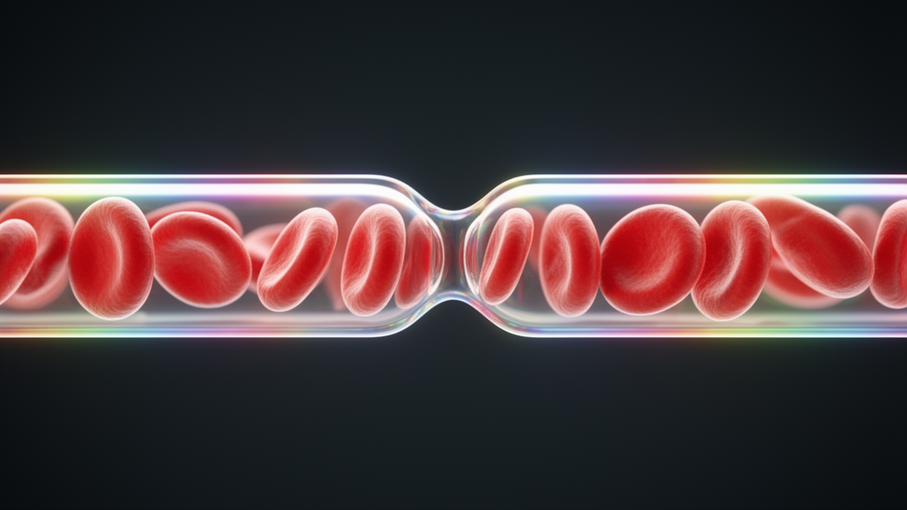
Clinicians currently judge these shapes by looking down a microscope, a method that works but depends heavily on the eye of the operator, resists reproducibility, and often demands fluorescent staining. A team reporting in Frontiers of Optoelectronics now describes a label-free alternative that reads the physical state of individual red blood cells from the way they twist the polarization of scattered laser light.
The study, led by Shuan Yao, Caizhong Guan, Nan Zeng and Honghui He, combines a dual-angle Mueller matrix polarimeter with computer simulations of polarized light scattering. A Mueller matrix is a sixteen-element mathematical object that fully characterizes how an object transforms the polarization state of light passing through or scattering off it. Rather than averaging signals over millions of cells in a suspension, as the group’s earlier work did, the new system isolates light from single cells one at a time as they drift through a focused laser beam, capturing polarization fingerprints that reveal size, shape, refractive index and even the presence of spiky membrane protrusions.
The instrument itself is a study in precision. A 473-nanometer solid-state laser feeds a polarization state generator built around a polarizer and two electro-optic modulators, which can switch the incident polarization state up to 25,000 times per second. A non-polarizing beam splitter divides the light: one path is focused to a spot smaller than 100 micrometers inside a dodecahedral glass sample chamber filled with refractive-index-matched water to suppress stray reflections, while the other serves as a reference. The chamber holds a stirred mouse red blood cell suspension, and as cells tumble randomly through the detection volume they produce brief scattered-light pulses. Two detection arms collect light simultaneously at 60 degrees, in the forward direction, and 120 degrees, in the backward direction, with pinholes constraining the scattering volume tightly enough to guarantee single-cell detection at concentrations below 100,000 cells per milliliter.
Behind the optics sits a four-quadrant polarization state analyzer that splits each scattered beam into four paths, measuring horizontal, vertical, 45-degree linear and circular polarization components with photomultiplier tubes. From these signals the full Stokes vector of the scattered light is reconstructed, and by cycling through many combinations of incident and scattered polarization states the system assembles the complete Mueller matrix at both angles. Calibration against standard polystyrene microspheres, whose reference matrices are calculated from Mie theory, corrects for polarization bias in the optical components and for non-polarization errors, keeping the measurements quantitatively trustworthy.
Measurement alone, however, does not explain what the numbers mean. To connect matrix elements to cell physics, the team turned to the discrete dipole approximation, a computational method that discretizes a cell’s volume into a grid of tiny dipoles and simulates how the whole ensemble scatters polarized light. Using a GPU-accelerated algorithm validated against Mie theory with errors below 0.01, they modeled biconcave red blood cells with realistic dimensions of six to nine micrometers, a relative refractive index near 1.053, and a wavelength matching the experiment. Because freely floating cells present every possible orientation to the beam, the simulations averaged results over many orientations, sampling thirty angles for symmetric shapes and a full 125-direction grid for spiculated models that lack symmetry.
The simulations revealed which matrix elements track which cell properties. At 60 degrees, elements such as M12 and M21 rise steadily with cell diameter, while M22 at 120 degrees falls; diagonal elements M33 and M44 at 60 degrees climb with refractive index, making them sensitive probes of hemoglobin concentration. Shape changes from flattened biconcave disc toward sphere produced monotonic shifts in five forward-scattering elements, and surface spiculation broke the reciprocity symmetry of the non-diagonal elements, with M12 and M21 at 120 degrees emerging as the cleanest indicators of spiky membranes. From these patterns the researchers distilled six polarization feature parameters with direct physical interpretations: two shape parameters K1 and K2 that approach zero as cells become spherical, a size parameter T120, a refractive index parameter T60, a surface parameter LE120 that flags spiculation, and a composite parameter LE60.
To test whether these parameters survive contact with real biology, the team stressed mouse red blood cells in three ways. Osmotic stress, delivered by NaCl solutions ranging from 150 to 800 milliosmoles, swelled cells in dilute conditions and shrank them in concentrated ones, with spiculation appearing above 600 milliosmoles. The polarization parameters responded exactly as the simulations predicted: T120 and LE60 rose with swelling and fell with shrinkage, T60 moved inversely with hemoglobin dilution and concentration, K1 and K2 tracked the loss and recovery of concavity, and LE120 stayed flat until spicules formed, then dropped sharply. Each box plot summarized 500 individual cells, giving the trends statistical weight.
Oxidative and acid-base stresses told a complementary story. Treating cells with hydrogen peroxide for one hour at 37 degrees Celsius induced spiculated shapes confirmed by optical microscopy, and the treated cells showed significantly increased K1 and K2 alongside decreased LE120 compared with phosphate-buffered saline controls, at p below 0.001 by t-test, while T120 changed only slightly and T60 not at all, indicating that oxidation reshaped the cells without much altering their size or refractive index. Acidic conditions at pH 6.8 swelled the cells and reduced concavity, raising T120 and LE60 while leaving LE120 stable, whereas alkaline conditions at pH 8.0 shrank cells, preserved concavity and produced marginal spiculation, with LE120 rising significantly. T60 remained constant across all pH groups, confirming that pH barely touched the intracellular refractive index.
The final demonstration pushed the method toward a practical screening task. The researchers mixed normal, spherical and spiculated cells in five known volume ratios and asked a random forest classifier, trained on 2,000 data points per cell type, to estimate the proportions of each type in the mixture. Using either the full 30-element Mueller matrix or just the six polarization feature parameters as inputs, the classifier quantified abnormal cell fractions with a maximum absolute error of 5.2 percent for the matrix-based model and 6.1 percent for the parameter-based model. The full matrix edged out the parameters in raw accuracy, but the parameters carried far clearer physical meaning, which the authors argue matters more for interpreting deformation mechanisms in real samples.
The work remains a proof of concept with clear boundaries: the experiments used mouse cells, the abnormal shapes were chemically induced in the laboratory rather than arising from human disease, and the spiculated cells were glutaraldehyde-fixed to hold their morphology stable during measurement. Even so, the combination of high-speed polarization switching, dual-angle single-cell detection and physically interpretable feature parameters sketches a pipeline that could one day flag pathological red blood cell populations quickly, without stains or subjective visual scoring.
Subject of Research: Label-free single-cell polarization measurement of red blood cell deformation using dual-angle Mueller matrix polarimetry
Article Title: Analysis of erythrocyte deformation characteristics based on dual-angle Mueller matrix measurement
Article References: Yao, S., Guan, C., Zeng, N., & He, H. (2025). Analysis of erythrocyte deformation characteristics based on dual-angle Mueller matrix measurement. Frontiers of Optoelectronics, 18(1), Article 23. https://doi.org/10.1007/s12200-025-00166-2
Image Credits: AI Generated
DOI: 10.1007/s12200-025-00166-2
Keywords: red blood cells, Mueller matrix polarimetry, polarized light scattering, discrete dipole approximation, polarization feature parameters, random forest classifier, osmotic stress, oxidative stress, pH stress, echinocytes, spherocytes, label-free cytometry
Cite Scienmag News
Denise Maddox. (October 28, 2026). Polarization Imaging Reads Deformed Red Blood Cells One Cell at a Time. Scienmag. https://scienmag.com/polarization-imaging-reads-deformed-red-blood-cells-one-cell-at-a-time/
Denise Maddox. "Polarization Imaging Reads Deformed Red Blood Cells One Cell at a Time." Scienmag, 28 October 2026, https://scienmag.com/polarization-imaging-reads-deformed-red-blood-cells-one-cell-at-a-time/. Accessed 28 October 2026.
Denise Maddox. "Polarization Imaging Reads Deformed Red Blood Cells One Cell at a Time." Scienmag. October 28, 2026. https://scienmag.com/polarization-imaging-reads-deformed-red-blood-cells-one-cell-at-a-time/


