


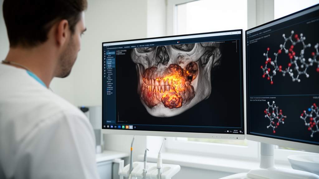
Artificial intelligence is moving steadily along the entire oral cancer care pathway, according to a narrative review published in Discover Artificial Intelligence, and the trajectory it describes is more ambitious than the familiar story of models that simply classify tissue images. The review, led by Shivam Agarwal and colleagues, maps how machine learning and deep learning now process clinical photographs, digitized histopathological slides, whole-slide images, radiological examinations, and molecular or clinical data to generate task-specific outputs, from the first identification of a suspicious oral lesion to the prediction of how an individual patient will respond to radiotherapy, chemotherapy, targeted therapy, or immunotherapy. The authors argue that the relationship between the input data, the computational task, and the clinically relevant output matters more than model architecture alone, and that this integrated perspective is what separates their synthesis from earlier literature focused on isolated diagnostic applications or headline performance figures.
The clinical problem the technology addresses is stark. Oral squamous cell carcinoma accounts for the majority of malignant tumors arising in the oral cavity, and the disease carries substantial morbidity and mortality largely because many patients are diagnosed at an advanced stage. This is particularly frustrating because the oral cavity is accessible to direct examination, yet early diagnosis remains difficult in routine practice. Oral carcinogenesis is a multistep process involving progressive genetic, molecular, and cellular changes in the oral epithelium, and these changes may appear clinically as oral potentially malignant disorders such as leukoplakia, erythroplakia, mixed red-white lesions, or persistent ulceration. On biopsy, such lesions may show hyperkeratosis, epithelial hyperplasia, or varying degrees of epithelial dysplasia. Crucially, not all potentially malignant disorders progress to invasive cancer, so the central diagnostic challenge is not merely to spot an abnormal lesion but to recognize the lesions with a higher risk of malignant transformation.
Conventional histopathological examination remains the reference standard for diagnosing epithelial dysplasia and oral squamous cell carcinoma, providing essential information on tumor differentiation, invasion, surgical margins, stromal response, and other prognostic features. Yet the review catalogues measurable limitations in this pathway. A biopsy samples only a selected area of a lesion and may not fully represent tissue heterogeneity, while the grading of dysplasia and the evaluation of tumor-related features can vary between observers, particularly in borderline or heterogeneous lesions. Studies of oral epithelial dysplasia have reported only fair-to-moderate interobserver agreement. Manual microscopy also remains limited in the consistent quantification of nuclear morphology, tissue organization, tumor-stroma interactions, immune-cell distribution, and spatial heterogeneity. These constraints can complicate early risk assessment and clinical decision-making, and they form the quantitative case for more objective and reproducible approaches.
Digital pathology is the bridge between visual assessment and computation. Whole-slide imaging converts complete histological sections into high-resolution digital images, organized as a multiresolution pyramid in which the highest-resolution level retains fine morphological detail while lower-resolution levels permit rapid navigation across the tissue. Unlike a static photomicrograph, which represents only a selected field, a whole-slide image preserves the spatial context of the tissue section and allows continuous navigation from low-power architecture to high-power cellular and nuclear detail. Direct comparisons have shown comparable diagnostic performance for whole-slide imaging versus conventional light microscopy, and oral pathology data indicate that a larger tissue area can be examined in less time without a significant increase in diagnostic discordance. Once digitized, the tissue section becomes a persistent image dataset that can be retrieved, annotated, measured, shared for remote consultation, and linked with laboratory and clinical information, providing the image substrate that computational analysis requires.
The review describes a distinct branch of AI operating before any biopsy is taken. Because the oral cavity is directly accessible to photography using smartphones, intraoral cameras, or conventional digital cameras, visible-light imaging offers a practical route for AI-assisted screening. The analytical pathway begins with image acquisition, followed by quality assessment to identify blur, inadequate illumination, flash reflection, poor framing, or incomplete visualization of the lesion. Detection or segmentation models then delineate the suspicious area, while classification models analyse visible characteristics including colour heterogeneity, white or red change, surface texture, ulceration, lesion border, shape, and spatial extent, mapping these representations to outputs such as a bounding box, segmentation mask, class probability, or a triage category of non-suspicious versus suspicious. The evidence shows that performance depends strongly on the diagnostic task: models generally perform better when distinguishing obvious established cancer from non-cancer images than when separating clinically overlapping early lesions, and performance is typically lower for leukoplakia, oral lichen planus, and other potentially malignant or benign lesions with overlapping appearances. A system that accurately recognizes established cancer does not necessarily provide equally reliable early-lesion screening.
Once H&E-stained tissue is digitized, AI can analyse microscopic morphology across selected image regions or the entire slide. The model can systematically search large tissue areas, assign probabilities to suspicious regions, and provide spatial information showing where malignant morphology is most strongly represented, with predictions mapped back to their original position to produce a heatmap or tumor map. Different architectures address different components: CNN-based classifiers such as ResNet and DenseNet learn local morphological patterns for discriminating malignant from non-malignant tissue, segmentation networks such as U-Net delineate the spatial extent of tumor regions, multiple-instance learning aggregates information from numerous tissue patches for slide-level diagnosis, and transformer-based models capture relationships between spatially separated regions. Recent hybrid CNN-transformer systems have reported striking internal results, including an explainable Vision Transformer framework achieving 98.45 percent accuracy with an AUC of 0.99 on a public histopathology dataset, and a hybrid ResNet50-ViT model reaching 98.97 percent accuracy on 5192 images. Publicly accessible datasets have supported accuracies of 99.30 percent and 96.66 percent for OSCC classification, though the review cautions that these estimates are not head-to-head comparisons because preprocessing, augmentation, partitioning, and validation differed between studies.
The review is emphatic that high internal accuracy should be interpreted cautiously. Models evaluated only on internally partitioned datasets may perform differently when applied to independent cohorts, and augmentation or multiple patches from the same patient do not increase the number of independent patients. A smaller group of studies has moved beyond internal validation: one evaluated oral epithelial dysplasia detection using an independent external dataset, while an explainable model for predicting malignant transformation in dysplasia using interpretable morphological and spatial features was independently validated in external cohorts from two centres, achieving an externally validated AUROC of 0.75 with a recall of 0.92, compared with an AUROC of 0.72 and a recall of 0.85 for binary dysplasia grading. A multiresolution pathology model developed using 221 whole-slide images achieved 80.0 percent accuracy, an F1-score of 77.3 percent, and an AUROC of 0.798 for predicting progression to invasive cancer. These findings indicate that computational pathology is moving beyond tissue classification toward risk stratification and outcome-oriented analysis.
AI-assisted grading of tumor differentiation remains less mature than binary cancer classification, constrained by limited datasets, variability in the pathological reference standard, and heterogeneous differentiation patterns within the same tumor. Its main potential value lies not in automated grade assignment but in more consistent assessment of heterogeneous morphology, since spatial mapping may reveal small poorly differentiated regions that could be under-represented when grading is based on a limited field. In progression and prognosis, AI can compare sequential CT, MRI, or PET examinations to quantify changes in tumor dimensions and volume, depth of local invasion, cervical lymph-node burden, and metastatic lesions, using image registration to align corresponding tumor regions across successive examinations. Pathomics-based studies have shown that prognostic information can be extracted directly from H&E-stained whole-slide images and combined with clinicopathological variables: a multicentre OSCC study using 240 whole-slide images demonstrated that supervised deep learning could predict overall survival and that integrating pathomics with clinical signatures further improved patient-level risk assessment.
In precision therapeutics, the role of AI shifts from disease recognition to treatment-specific decision support. Patients with apparently similar tumor sites, stages, and histopathological characteristics may show substantially different responses to therapy, and therapeutic heterogeneity arises from differences in tumor architecture, molecular alterations, immune composition, intratumoral heterogeneity, and microenvironmental interactions. Models learn the relationship between pretreatment characteristics and subsequently observed outcomes, integrating histopathological, radiological, molecular, and clinical data to estimate a treatment-specific probability of response, resistance, or benefit, supporting selection, intensification, de-escalation, or modification of therapy. However, the review notes that evidence for AI-assisted precision therapeutics in oral cancer remains relatively limited, with several proposed approaches informed by broader head-and-neck or pan-cancer evidence, and that multimodal models’ added clinical value over established clinicopathological assessment requires oral cancer-specific external and prospective validation.
The authors are candid about the barriers between promising performance and routine practice. Major challenges include dataset quality and representativeness, domain shift across scanners and staining protocols, annotation variability, interpretability, and integration into clinical workflows. Explainable AI can identify image regions contributing to a prediction, but a plausible heatmap does not establish that a model will remain accurate across different laboratories, scanners, populations, or clinical settings; model justification requires agreement between highlighted regions and established pathological features, expert review, robustness testing, independent external validation, and assessment of predictive uncertainty. On the review’s own translational scale, most oral cancer AI studies currently sit between development and retrospective external validation, while prospective workflow and clinical-impact evaluation remain the important next steps. Future progress will require large multicentre datasets including early lesions and diagnostically difficult mimics, patient-level evaluation, multimodal integration, uncertainty-aware prediction, foundation models and self-supervised learning validated specifically for oral lesions, federated learning for privacy-preserving multicentre development, and continuous monitoring after deployment. The long-term objective, the authors stress, is not an autonomous system that replaces clinical or pathological expertise, but an integrated human-AI framework in which computational models support lesion triage, diagnosis, grading, longitudinal disease assessment, and individualized treatment selection across the oral cancer care pathway.
Subject of Research: Artificial Intelligence in Medicine
Article Title: AI driven oral cancer diagnosis from clinical imaging and computational pathology to precision therapeutics
Article References: Agarwal, S., Das, B., Singh, S., & Gupta, B. P. (2026). AI driven oral cancer diagnosis from clinical imaging and computational pathology to precision therapeutics. Discover Artificial Intelligence, 6(1), Article 1611. https://doi.org/10.1007/s44163-026-02573-6
Image Credits: AI Generated
DOI: 10.1007/s44163-026-02573-6
Keywords: artificial intelligence, oral cancer, computational pathology, whole-slide imaging, precision oncology, driven, oral, cancer, diagnosis, clinical, imaging, computational
Cite Scienmag News
Nathaniel Bowman. (October 25, 2026). AI reshapes oral cancer diagnosis from clinical imaging to precision therapy. Scienmag. https://scienmag.com/ai-reshapes-oral-cancer-diagnosis-from-clinical-imaging-to-precision-therapy/
Nathaniel Bowman. "AI reshapes oral cancer diagnosis from clinical imaging to precision therapy." Scienmag, 25 October 2026, https://scienmag.com/ai-reshapes-oral-cancer-diagnosis-from-clinical-imaging-to-precision-therapy/. Accessed 25 October 2026.
Nathaniel Bowman. "AI reshapes oral cancer diagnosis from clinical imaging to precision therapy." Scienmag. October 25, 2026. https://scienmag.com/ai-reshapes-oral-cancer-diagnosis-from-clinical-imaging-to-precision-therapy/


