


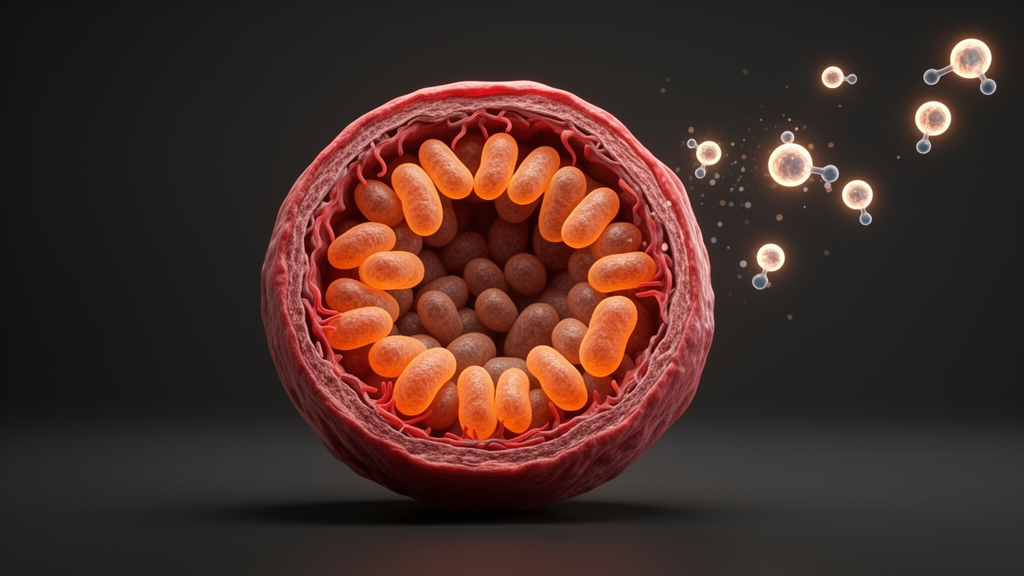
Cardiovascular disease is the leading cause of illness and death in women, yet the risk is not distributed evenly across a woman’s life. Before menopause, women carry roughly a 50 percent lower risk of cardiovascular disease than age-matched men, but that protection erodes as ovarian hormone production falters. A new review synthesizes evidence for a specific molecular explanation: estradiol, the predominant estrogen of the reproductive years, appears to sustain the health of mitochondria in heart muscle and blood vessel walls by regulating a family of transcriptional coactivators known as the PGC-1 family. As estradiol becomes erratic during perimenopause and then chronically low after menopause, the review proposes, this estradiol–PGC-1 signaling axis may weaken, compromising the energy factories of cardiovascular cells and helping to drive the rise in heart disease risk that follows the menopause transition.
The review, published in the journal Physiological Reports, frames mitochondrial dysfunction as a central mechanism in cardiovascular disease. Mitochondria are the organelles that generate most of a cell’s ATP through oxidative phosphorylation, and when they fail to meet cellular energy demands, several problems follow: ATP synthesis declines, reactive oxygen species accumulate, and the resulting oxidative burden damages cellular components. Within blood vessels, excess reactive oxygen species react with and deplete nitric oxide, the molecule that endothelial cells release to relax arteries. Less nitric oxide means impaired vasodilation, reduced blood flow, and greater cardiovascular strain—a pathological cycle that compounds over years and elevates long-term disease risk.
At the center of the proposed mechanism sits PGC-1α, widely regarded as the master regulator of mitochondrial biogenesis, alongside its relatives PGC-1β and PGC-1-related coactivator. These coactivators regulate the mitochondrial quality of skeletal muscle, cardiac muscle, and the vascular wall, coordinating programs of gene expression that build and maintain mitochondria in these tissues. Experimental work shows that when PGC-1α or PGC-1β expression falls, mitochondrial function deteriorates, oxidative stress resilience drops, and adaptive responses blunt. Conversely, forcing PGC-1α expression in cardiac muscle increases mitochondrial biogenesis, oxidative phosphorylation gene expression, fatty acid oxidation, and coupled respiration, while losing PGC-1α produces the opposite phenotype and impairs cardiac and metabolic responses.
Estradiol has long been described as cardioprotective: it increases endothelial nitric oxide synthase activity and nitric oxide–mediated vasodilation, enhances antioxidant and anti-inflammatory defenses, and promotes angiogenesis and anti-atherogenic signaling. The review’s key contribution is to connect these well-known effects to PGC-1 regulation through several distinct signaling routes. In the classical genomic pathway, estradiol diffuses across the cell membrane and binds intracellular estrogen receptors ERα or ERβ, which dimerize and bind estrogen response elements in the regulatory regions of target genes. PGC-1α and PGC-1β act as coactivators of these receptors—their LXXLL motifs engage the receptors’ activation function-2 domain—helping assemble transcriptional complexes that switch on genes for mitochondrial biogenesis such as NRF-1 and TFAM, mitochondrial DNA–encoded respiratory genes, nuclear-encoded oxidative phosphorylation genes, and antioxidant defenses like SOD2.
Beyond this direct route, the review details a tethered genomic mechanism in which ligand-bound estrogen receptors interact with transcription factors already bound to DNA—activator protein-1, NF-κB, and Sp1—thereby modulating stress-response, inflammatory, and tissue-specific gene programs without directly binding estrogen response elements. There is also rapid, non-genomic signaling: membrane-associated estrogen receptors, including palmitoylated ERα targeted to caveolae, trigger kinase cascades within seconds to minutes. These include p38 MAPK, a positive regulator of PGC-1α expression and activity, and PI3K/Akt signaling, which promotes eNOS phosphorylation and nitric oxide production while regulating glucose and lipid metabolism and intracellular calcium handling—elevated calcium activating CaMK and calcineurin pathways that can further promote PGC-1α expression.
A third route runs through GPER, the G protein–coupled estrogen receptor, which is largely intracellular but remains accessible to estrogen. Upon ligand binding, GPER activates Src family kinases, which trigger matrix metalloproteinase–mediated release of heparin-binding epidermal growth factor, transactivating the epidermal growth factor receptor and downstream cascades including p38 MAPK, PI3K/Akt, ERK1/2, AMPK, and cAMP/PKA. Two of these are especially relevant to mitochondrial regulation: AMPK can activate PGC-1α through phosphorylation, and SIRT1-mediated deacetylation further increases PGC-1α activity. In ovariectomized mice treated with 17β-estradiol, estradiol engaged an ERα–SIRT1 pathway that deacetylated and activated PGC-1α in liver, preserving mitochondrial function after ovarian hormone loss.
Human evidence, though more limited, is consistent with these mechanisms. Women within six years of menopause who received short-term, high-dose transdermal 17β-estradiol showed increased nuclear skeletal muscle PGC-1α abundance—about 22 percent—and greater AMPK phosphorylation. Notably, this responsiveness appeared in early but not late postmenopausal women, suggesting a window of timing dependence. Separately, long-term users of combined estradiol–progestin therapy exhibited greater skeletal muscle mitochondrial content and respiratory capacity, along with lower visceral and total fat mass, compared with matched nonusers. In young adult men, estradiol supplementation increased PGC-1α mRNA by 29 percent, hinting that the pathway is not exclusive to females. In rodents, chronic selective ERα activation in aged, high-fat-fed ovariectomized mice increased cardiac PGC-1α and NRF-1 expression, improved mitochondrial respiration, and enhanced insulin sensitivity and adiposity measures.
The review is careful to note that physiological estradiol fluctuations in healthy women produce more modest and context-dependent effects than the dramatic changes seen in experimental models. Studies comparing menstrual cycle phases have yielded mixed results: one reported reduced phosphocreatine recovery and ATP generation during the low-hormone early follicular phase compared with the high-estradiol late follicular phase, while another found no differences in maximal mitochondrial oxidative capacity or citrate synthase activity between phases, detecting only subtle changes in proton leak respiration and reactive oxygen species emission. Findings on substrate use during exercise across hormone phases are similarly contested. The clearest signal emerges with the menopause transition itself, when estradiol fluctuations become erratic—beginning on average four to seven years before the final menstrual period—and progesterone becomes variable, contributing to anovulatory cycles. Some data suggest progesterone increases mitochondrial reactive oxygen species emission regardless of estradiol availability and reduces respiration when estradiol is low, potentially compounding oxidative stress as cycles falter.
Cell-specific details add texture to the picture. Cardiomyocytes predominantly express full-length ERα66, while human endothelial cells are rich in the truncated ERα46, which efficiently mediates rapid, membrane-initiated vascular signaling through PI3K/Akt and MAPK to activate eNOS and nitric oxide production. Endothelial ERα expression is approximately 33 percent lower in postmenopausal women than in premenopausal women during the late follicular phase, and this reduction is associated with diminished vasodilatory capacity and greater susceptibility to atherosclerotic vascular dysfunction. In mouse models of heart failure, the PGC-1α promoter contains an ERα-binding site within its first 600 base pairs, and estradiol significantly increased PGC-1α promoter activity only in the presence of ERα—underscoring that sufficient receptor expression, not merely higher estradiol, is required for the genomic signaling that maintains mitochondrial gene programs. Female cardiomyocytes also show greater mitochondrial resilience than male cells, with more efficient respiratory chain organization, lower reactive oxygen species, and better calcium handling.
The review’s authors conclude that declining estradiol during the menopause transition may impair mitochondrial function by reducing pathways that support PGC-1 signaling, and that late perimenopause through early postmenopause represents a critical window of low estradiol availability that may raise cardiovascular risk in part through this mechanism. They also address the contested history of hormone therapy: after the 2002 Women’s Health Initiative trial linked hormone use to increased risks of breast cancer, stroke, coronary heart disease, and venous thromboembolism, US menopausal hormone therapy use dropped by 80 percent. More recently, a longitudinal cohort study of 876,805 women reported no increased risk of all-cause mortality with menopausal hormone therapy use, a discrepancy likely explained by the WHI participants being ten or more years postmenopause with an average age of 63 and many existing cardiometabolic diagnoses. The authors call for future studies to determine whether estradiol loss alters PGC-1–mediated cardiovascular pathways in an isoform-specific manner, and whether hormonal or non-hormonal strategies—such as exercise, which restored skeletal muscle PGC-1α in ovariectomized mice through AMPK/SIRT1 signaling independent of estradiol—can preserve mitochondrial function despite declining ovarian hormones. Direct measurements of estradiol–ERα signaling in human cardiomyocytes and endothelial cells across perimenopause remain limited, and the review’s proposals are framed as mechanisms to be tested rather than established clinical benefits.
Subject of Research: Estradiol regulation of PGC-1 coactivator signaling and cardiovascular mitochondrial health across the menopause transition
Article Title: Estradiol—PGC‐1 signaling in mitochondrial health: Implications for perimenopause and cardiovascular disease
Article References: Parten, A. L., & Rowe, G. C. (2026). Estradiol— PGC ‐1 signaling in mitochondrial health: Implications for perimenopause and cardiovascular disease. Physiological Reports, 14(20), Article e71155. https://doi.org/10.14814/phy2.71155
Image Credits: AI Generated
DOI: 10.14814/phy2.71155
Keywords: estradiol, PGC-1alpha, mitochondria, menopause, cardiovascular disease, estrogen receptors, perimenopause, hormone therapy, endothelial function, oxidative phosphorylation, GPER, women's health
Cite Scienmag News
Drew Townsend. (October 25, 2026). How Falling Estrogen May Weaken the Heart’s Mitochondria After Menopause. Scienmag. https://scienmag.com/how-falling-estrogen-may-weaken-the-hearts-mitochondria-after-menopause/
Drew Townsend. "How Falling Estrogen May Weaken the Heart’s Mitochondria After Menopause." Scienmag, 25 October 2026, https://scienmag.com/how-falling-estrogen-may-weaken-the-hearts-mitochondria-after-menopause/. Accessed 25 October 2026.
Drew Townsend. "How Falling Estrogen May Weaken the Heart’s Mitochondria After Menopause." Scienmag. October 25, 2026. https://scienmag.com/how-falling-estrogen-may-weaken-the-hearts-mitochondria-after-menopause/


