


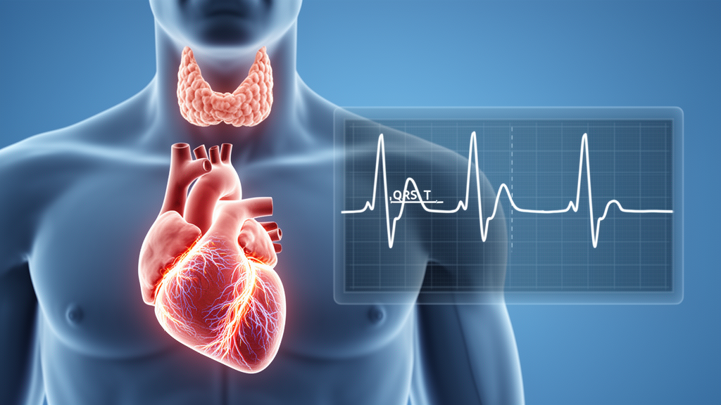
A retrospective, single-center study found that adults with Graves’ disease and biochemically confirmed overt hyperthyroidism had a wider frontal QRS-T angle than euthyroid controls. The study included 55 patients with overt hyperthyroidism and 42 controls. Their median frontal QRS-T angles were 21.89 degrees and 17.36 degrees, respectively, a statistically significant difference with a p-value of 0.006. The angle represents the directional relationship between ventricular depolarization and repolarization, and its wider value in the hyperthyroid group was consistent with greater discordance between those processes. However, the measure had only modest ability to distinguish the groups, with an area under the receiver operating characteristic curve of 0.664 and a 95% confidence interval of 0.552 to 0.768.
The research examined 12-lead electrocardiograms and laboratory data from people admitted to a tertiary referral center in southeastern Poland between June 1, 2022, and June 30, 2025. Patients were eligible if they were 18 years or older, had a documented history of hyperthyroidism due to Graves’ disease, and had overt hyperthyroidism, defined biochemically as suppressed thyroid-stimulating hormone, or TSH, together with free thyroxine, or FT4, and/or free triiodothyronine, or FT3, above the relevant laboratory reference ranges. Of 953 potentially eligible records identified during screening, 55 met all eligibility requirements and entered the hyperthyroid group. Controls were people whose hospital visits were unrelated to thyroid or cardiac disease and whose TSH, FT3 and FT4 concentrations all fell within their respective reference ranges. They also had no history of thyroid disease and met the same restrictions on cardiovascular illness, systemic disorders and medications as the hyperthyroid group.
Exclusion criteria were intended to reduce alternative explanations for electrocardiographic differences. The researchers excluded people with coronary artery disease, previous myocardial infarction, heart failure, cardiomyopathy, clinically significant valvular or congenital heart disease, clinically significant arrhythmias and cardiac conduction disorders. They also excluded electrolyte imbalances, significant endocrine, renal, hepatic, inflammatory or other systemic conditions, and medications that could affect myocardial function or cardiac electrophysiology. Relevant excluded medicines included antiarrhythmics, beta-blockers, non-dihydropyridine calcium-channel blockers, cardiac glycosides, sympathomimetic or sympatholytic agents and QT-prolonging drugs. The resulting groups were comparable in age, sex distribution and body mass index, with all comparisons for those characteristics yielding p-values above 0.05. The study population was nevertheless small, and its retrospective design limited the ability to assess causal relationships, changes over time or reversibility after thyroid function improved.
Each electrocardiogram was a standard 10-second, 12-lead recording obtained at a paper speed of 25 millimeters per second and an amplitude calibration of 10 millimeters per millivolt. When several tracings were available for a patient, investigators selected the one closest in time to the blood sample used to assess thyroid function. They then measured QRS-complex and T-wave amplitudes in leads I and aVF with ImageJ, using the isoelectric line as the reference level. Net QRS amplitude was calculated by algebraically summing positive and negative deflections. T-wave amplitude was measured from the isoelectric line to the peak, with negative deflections assigned the corresponding sign. Measurements from three consecutive cardiac cycles were averaged for each tracing.
The frontal electrical axes of the QRS complex and T wave were calculated from the measured amplitudes in leads I and aVF with a two-argument arctangent function. The absolute difference between those axes gave the raw angular separation. When that separation exceeded 180 degrees, 360 degrees was subtracted from it, producing a final frontal QRS-T angle between 0 and 180 degrees. A larger angle represents greater directional discordance between ventricular depolarization and repolarization and may reflect less uniform ventricular electrical behavior. The key analytical finding was that the hyperthyroid and control groups did not differ significantly in their QRS axes or T-wave axes considered separately. The investigators therefore interpreted the wider combined angle as reflecting an altered relationship between the two electrical processes rather than a uniform shift in the direction of either one.
The groups differed markedly in heart rate and thyroid hormone status. Median heart rate was 102 beats per minute, with an interquartile range of 94 to 113.5, among patients with overt hyperthyroidism, compared with 74.4 beats per minute, with an interquartile range of 65.9 to 85.1, among controls; the difference had a p-value below 0.001. Median FT3 concentrations were 8.52 pg/mL in the hyperthyroid group and 2.95 pg/mL in controls, while median FT4 concentrations were 3.28 ng/dL and 1.10 ng/dL, respectively. Median TSH was 0.007 µIU/mL in the hyperthyroid group and 1.74 µIU/mL in controls. All three thyroid-function comparisons had p-values below 0.001. N-terminal pro-B-type natriuretic peptide and high-sensitivity cardiac troponin T were also higher in the hyperthyroid group, with p-values of 0.003 and below 0.001, respectively, although these biomarkers were available only in subsets of participants.
Neither of the individual frontal axes differed significantly between groups. The median QRS axis was 35.5 degrees, with an interquartile range of 14.7 to 61.6 degrees, in the hyperthyroid group and 39.0 degrees, with an interquartile range of 12.4 to 55.4 degrees, in controls, producing a p-value of 0.774. Median T-axis values were 32.4 and 32.6 degrees, with p = 0.675. Circular statistical analyses also found no significant differences in the mean direction of either axis, with both p-values above 0.70. The primary comparison nevertheless showed a modest effect size expressed as a rank-biserial correlation of 0.328, with a bootstrap 95% confidence interval of 0.106 to 0.535. Thirteen of the 55 patients with overt hyperthyroidism, or 23.6%, had an angle above 45 degrees, while none of the 42 controls exceeded that threshold; Fisher’s exact p-value was below 0.001. Angles above 90 degrees were uncommon, appearing in only one hyperthyroid patient and no controls, with no significant between-group difference.
Because the faster heart rate in the hyperthyroid group could have influenced the result, the investigators fitted a multivariable model that included overt hyperthyroidism, age, sex, body mass index and heart rate. After adjustment, overt hyperthyroidism remained associated with a 9.00-degree greater frontal QRS-T angle, with a 95% confidence interval of 1.77 to 16.22 degrees and a p-value of 0.015. Heart rate was not independently associated with the angle in that model, with a coefficient of 0.132 degrees per beat per minute and p = 0.170. There was also no significant interaction between hyperthyroidism status and heart rate, with p = 0.479. Sensitivity analyses supported the main result: after logarithmic transformation, the geometric mean ratio was 1.68, with a 95% confidence interval of 1.10 to 2.54 and p = 0.015, while a generalized linear model with a gamma distribution and log link produced a ratio of 1.58, a 95% confidence interval of 1.14 to 2.18 and p = 0.006. These models indicate that the observed group difference was not explained solely by the increased heart rate accompanying hyperthyroidism.
Exploratory analyses did not identify a clear relationship between the angle and the biochemical severity of hyperthyroidism. Within the hyperthyroid group, the frontal QRS-T angle was not significantly correlated with FT3, FT4, TSH or heart rate. The respective Spearman correlation coefficients were 0.129, 0.243, −0.169 and 0.136, with p-values of 0.348, 0.074, 0.218 and 0.321. None remained significant after correction for multiple comparisons. No measured renal-function or electrolyte variable was significantly related to the angle: these analyses included creatinine, estimated glomeronal filtration rate, sodium, potassium, chloride and calcium. The authors described these exploratory findings cautiously because the cross-sectional analysis measured hormone concentrations and electrocardiograms at one point and could not determine whether the relationship changed with cumulative exposure or treatment. The angle should not be interpreted as a diagnostic marker of overt hyperthyroidism, given its modest discrimination and lack of external threshold validation.
Measurement reproducibility was strong. Investigators repeated the axis and angle measurements under identical conditions in a randomly selected subset of 60 participants, comprising 30 patients with overt hyperthyroidism and 30 controls. Intraclass correlation coefficients for absolute agreement were 0.97 for the QRS axis, with a 95% confidence interval of 0.94 to 0.99; 0.96 for the T axis, with a 95% confidence interval of 0.92 to 0.98; and 0.94 for the frontal QRS-T angle, with a 95% confidence interval of 0.88 to 0.97. The study nevertheless has important limitations: it was retrospective, single-center and relatively small; the duration of overt hyperthyroidism before hospitalization was unknown; and residual confounding could not be excluded despite statistical adjustment. Controls were emergency-department patients selected retrospectively rather than a community-based reference population, so acute illness, transient physiological disturbances or the reasons for their visits may have affected electrocardiographic measurements. Prospective, multicenter and longitudinal studies with repeated electrocardiograms before and after restoration of euthyroidism are needed to determine whether the observed widening is reversible and whether it has prognostic significance for arrhythmias or cardiovascular outcomes.
Subject of Research: Association of overt hyperthyroidism in Graves’ disease with frontal QRS-T angle on electrocardiography
Article Title: Frontal QRS-T angle in patients with Graves’ disease: association with thyroid function and cardiovascular parameters
Article References: Kłosowicz, M., Urbańczuk, M., Burbelka, A., Gala-Błądzińska, A., & Balawender, K. (2026). Frontal QRS-T angle in patients with Graves’ disease: association with thyroid function and cardiovascular parameters. Frontiers in Endocrinology, 17, 1979248. https://doi.org/10.3389/fendo.2026.1979248
Image Credits: AI Generated
DOI: 10.3389/fendo.2026.1979248
Keywords: Graves’ disease, overt hyperthyroidism, frontal QRS-T angle, electrocardiography, ventricular depolarization, ventricular repolarization, cardiac electrophysiology, thyroid hormone, heart rate, Frontal, QRS-T, angle
Cite Scienmag News
Frances Kline. (October 22, 2026). Graves’ hyperthyroidism linked to wider ventricular QRS-T angle. Scienmag. https://scienmag.com/graves-hyperthyroidism-linked-to-wider-ventricular-qrs-t-angle/
Frances Kline. "Graves’ hyperthyroidism linked to wider ventricular QRS-T angle." Scienmag, 22 October 2026, https://scienmag.com/graves-hyperthyroidism-linked-to-wider-ventricular-qrs-t-angle/. Accessed 22 October 2026.
Frances Kline. "Graves’ hyperthyroidism linked to wider ventricular QRS-T angle." Scienmag. October 22, 2026. https://scienmag.com/graves-hyperthyroidism-linked-to-wider-ventricular-qrs-t-angle/


