


A retrospective cohort study of 500 preterm infants found that a nine-factor nomogram combining three blood markers of inflammation with five abdominal ultrasound measurements predicted early-stage neonatal necrotizing enterocolitis with good discrimination and calibration. The model achieved an area under the receiver operating characteristic curve of 0.880 in a development cohort of 350 infants and 0.850 in an internal validation cohort of 150. At a fixed predicted-risk threshold of 0.18, sensitivity was 82.4% and specificity 81.2% in development, compared with 77.2% and 76.0% in validation. The findings support further evaluation of the nomogram as a noninvasive screening aid, but the single-center, retrospective study did not establish that using it improves clinical outcomes.
Neonatal necrotizing enterocolitis, commonly called NEC, is the most common severe gastrointestinal emergency among preterm and very-low-birth-weight infants, according to the study’s background. It involves bowel-wall ischemia, inflammation and necrosis, often has nonspecific early symptoms and can progress rapidly. Conventional abdominal radiography may not reveal subtle bowel-wall changes during early disease. Abdominal ultrasound is noninvasive, repeatable and free of radiation, and it can display bowel-wall thickness, intestinal movement, blood perfusion and subtle intra-abdominal changes. The researchers therefore tested whether a visual prediction model combining laboratory inflammation markers with multiparametric ultrasound could quantify an infant’s risk of early NEC.
The participants were 500 infants born before 37 weeks’ gestation who had been admitted to the neonatal intensive care unit of Quanzhou Maternal and Child Health Hospital in China within 24 hours of birth. Eligibility also required complete clinical, laboratory and ultrasound data. The investigators excluded infants with congenital gastrointestinal malformations, congenital heart disease or inherited metabolic diseases, as well as those with severe birth asphyxia, severe intracranial hemorrhage or surgery after admission. Stays shorter than 72 hours and loss to follow-up were also excluded. NEC was diagnosed using modified Bell staging criteria, with clinical findings, abdominal ultrasound and radiographic examinations contributing to the determination. All cases were stage I or II; severe stage III disease was excluded, making the model specific to early disease.
Within the retrospective cohort, infants were stratified by whether they developed NEC and then randomly allocated in a 7:3 ratio to development and validation groups. The development cohort contained 350 infants, including 68 who developed NEC and 282 who did not. The validation cohort contained 150 infants, of whom 29 developed NEC and 121 did not. Altogether, 97 of the 500 infants developed early-stage NEC. The two cohorts were internal subsets of the same single-center sample rather than independent populations recruited from different hospitals. Clinical variables were taken from electronic medical records, and C-reactive protein, procalcitonin and interleukin-6 were measured in peripheral venous blood collected within 72 hours of admission.
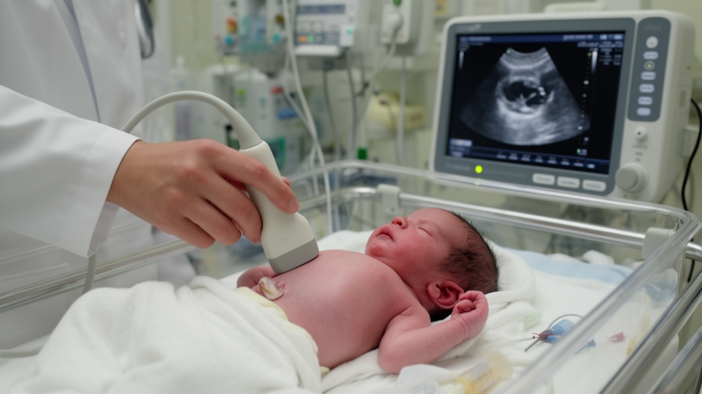
Two senior ultrasonographers performed color Doppler examinations without access to clinical manifestations or laboratory results. They used a probe frequency of 7.5 to 10.0 MHz and recorded bowel-wall thickness, intestinal peristalsis, gastric antral area, gastric antral peristalsis and bowel-wall blood perfusion. Bowel-wall thickening was defined as a thickness of at least 2 mm, reduced intestinal peristalsis as fewer than three movements per minute, enlarged gastric antral area as more than 1.5 cm², and reduced gastric antral peristalsis as fewer than two movements per minute. Intramural blood flow was categorized as normal, sparse, absent or overperfused, with the latter three grouped as abnormal for modeling. Inter-rater reliability was assessed in 80 randomly selected cases. Intraclass correlation coefficients were 0.84 for bowel-wall thickness and 0.81 for gastric antral area, while Cohen’s kappa values ranged from 0.76 to 0.80 for the categorical ultrasound measures.
In the development cohort, infants who developed NEC had a mean gestational age of 31.17 ± 2.21 weeks, compared with 34.94 ± 2.03 weeks among those who did not. Mean birth weight was 1,442.74 ± 364.21 grams in the NEC group and 2,197.43 ± 428.17 grams in the non-NEC group. Mean C-reactive protein was 18.81 ± 5.47 mg/L versus 6.14 ± 2.20 mg/L; mean procalcitonin was 3.94 ± 1.31 ng/mL versus 0.82 ± 0.33 ng/mL; and mean interleukin-6 was 158.74 ± 47.13 pg/mL versus 57.41 ± 18.62 pg/mL. Abnormal bowel-wall perfusion occurred in 94.12% of infants with NEC and 19.15% of those without it. Sex, delivery mode and Apgar scores did not differ significantly between the groups in the univariate analysis.
Multivariable logistic regression identified nine independent associations with early NEC: gestational age, C-reactive protein, procalcitonin, interleukin-6, bowel-wall thickening, reduced intestinal peristalsis, enlarged gastric antral area, reduced gastric antral peristalsis and abnormal bowel-wall perfusion. Birth weight was associated with NEC in the univariate analysis but was excluded from the final model because of strong collinearity with gestational age; their variance inflation factors were 7.82 and 8.14. The nine retained predictors had variance inflation factors between 1.21 and 1.48. Each additional week of gestational age was associated with lower odds of early NEC, with an odds ratio of 0.831 and 95% confidence interval of 0.765–0.902. The odds ratios for the three laboratory markers were 1.242 for C-reactive protein, 1.385 for procalcitonin and 1.219 for interleukin-6.
The five ultrasound variables also remained associated with early NEC after adjustment. The model’s odds ratio was 1.572 for bowel-wall thickening, 8.215 for reduced intestinal peristalsis, 2.452 for enlarged gastric antral area, 7.265 for reduced gastric antral peristalsis and 14.456 for abnormal bowel-wall perfusion. The researchers converted the nine regression coefficients into a nomogram with separate scoring axes, a total-score axis and a risk axis for early NEC. Although the tool is described as ultrasound-based, it also requires gestational age and three laboratory measurements, making it a multimodal model rather than a stand-alone imaging test. Sensitivity analyses that removed one inflammatory marker at a time left the direction and statistical significance of the remaining associations unchanged, while the development-cohort AUC varied only between 0.861 and 0.883.
When the Youden index was used in the development cohort, 0.18 was selected as the optimal predicted-risk threshold and then applied without adjustment to the validation cohort. At this cutoff, the development cohort produced 82.4% sensitivity, 81.2% specificity, 51.4% positive predictive value and 95.0% negative predictive value. In validation, the corresponding measures were 77.2%, 76.0%, 43.8% and 92.4%. Thus, the positive predictive value was lower in validation: among infants flagged as positive, fewer than half developed early NEC in that cohort. By contrast, a negative result was associated with a high probability that early NEC did not occur during follow-up. Calibration was favorable, with goodness-of-fit chi-square values of 1.235 and P = 0.998 in development and 1.642 and P = 0.995 in validation. Decision-curve analysis suggested net benefit over treating all or none when the probability threshold exceeded 0.05.
Several limitations constrain the findings. As a retrospective, single-center study, the model is susceptible to selection bias and may not generalize to hospitals with different patient populations, resources or ultrasound practices. Although the investigators planned for at least 10 NEC events per predictor, the development cohort contained approximately 7.5 events per predictor, below the conventional logistic-regression benchmark. Feeding patterns, intestinal microbiota and umbilical arterial catheterization were not included, and no formal correction for multiple comparisons was applied during univariate screening. Stage I cases represent suspected NEC with limited diagnostic specificity, which may have affected the estimates. Follow-up lasted 28 days and could therefore miss later events, while ultrasound measurements remain partly operator-dependent and were assessed for inter-rater reliability in only 80 infants. Sparse, absent and hyperperfused perfusion were also collapsed into one binary category.
Overall, the study shows that gestational age, inflammatory markers and multiparametric ultrasound findings can be combined into a quantitative model with favorable internal predictive performance for early-stage NEC in preterm infants. The lower validation positive predictive value indicates that the tool should not be interpreted as a definitive diagnosis, and the research did not test whether nomogram-guided care reduces missed diagnoses, disease progression or mortality. Prospective, multicenter external validation is still needed to establish performance in other neonatal intensive care units and to determine whether the model can support decisions in practice. Until then, the results are best viewed as evidence that bedside ultrasound and conventional laboratory information can complement one another in neonatal risk assessment, not as proof of a new diagnostic standard.
Subject of Research: Internal development and validation of a nomogram predicting early-stage neonatal necrotizing enterocolitis in preterm infants using inflammatory markers and abdominal ultrasound.
Article Title: An ultrasound-based nomogram for predicting early neonatal necrotizing enterocolitis
Article References: RUAN, J., SU, H., HE, Y., & LIN, W. (2026). An ultrasound-based nomogram for predicting early neonatal necrotizing enterocolitis. Frontiers in Pediatrics, 14, Article 1907406. https://doi.org/10.3389/fped.2026.1907406
Image Credits: AI Generated
DOI: 10.3389/fped.2026.1907406
Keywords: neonatal necrotizing enterocolitis, preterm infants, abdominal ultrasound, clinical nomogram, risk prediction, inflammation markers, bowel perfusion, internal validation, neonatal intensive care, ultrasound-based, nomogram, predicting
Cite Scienmag News
Harold Sullivan. (October 20, 2026). An ultrasound-based nomogram for predicting early neonatal necrotizing enterocolitis. Scienmag. https://scienmag.com/an-ultrasound-based-nomogram-for-predicting-early-neonatal-necrotizing-enterocolitis/
Harold Sullivan. "An ultrasound-based nomogram for predicting early neonatal necrotizing enterocolitis." Scienmag, 20 October 2026, https://scienmag.com/an-ultrasound-based-nomogram-for-predicting-early-neonatal-necrotizing-enterocolitis/. Accessed 20 October 2026.
Harold Sullivan. "An ultrasound-based nomogram for predicting early neonatal necrotizing enterocolitis." Scienmag. October 20, 2026. https://scienmag.com/an-ultrasound-based-nomogram-for-predicting-early-neonatal-necrotizing-enterocolitis/


