


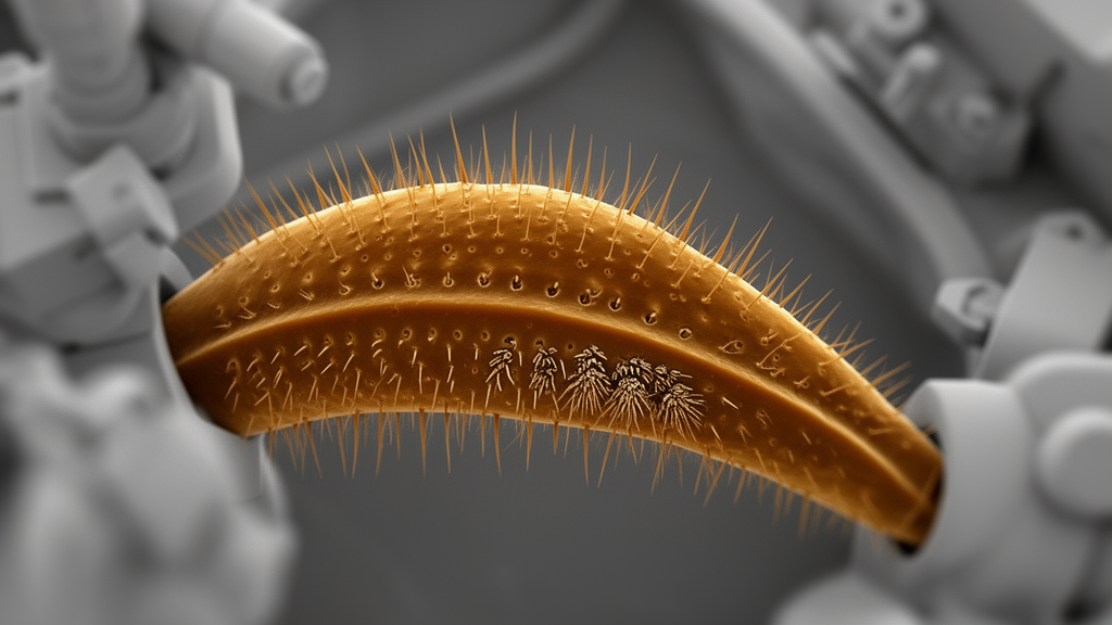
Scanning electron microscopy of mouthpart palps from 25 caddisfly species representing seven basal families identified seven types of sensilla and showed that their distribution is broadly conservative across lineages, despite family-level and species-level modifications. The study also compared two basal moths as external outgroups and found a closely corresponding core set of sensory structures. The results support a hypothetical ancestral arrangement for caddisfly palps, while also documenting the repeated reduction or loss of particular structures in different branches.
The researchers examined caddisflies in the Rhyacophilidae, Glossosomatidae, Hydroptilidae, Ptilocolepidae, Hydrobiosidae, Philopotamidae and Stenopsychidae. Most specimens came from the collection of the Department of Entomology at St. Petersburg State University and had been preserved in 70% ethanol.
The sample spanned multiple genera and included males and females for some species. Two basal Lepidoptera, Micropterix maschukella and Eriocrania cicatricella, were included for comparison. This was a comparative morphology study based on the external structure and arrangement of receptors; it did not measure neural activity or test behavior experimentally.
For scanning electron microscopy, the researchers removed the palps or heads, dried them, mounted them on specimen holders and coated them with 20 nm of gold. They used three scanning electron microscopes and counted and measured sensilla from the resulting micrographs with ImageJ 1.52r. The insects had three-segmented labial palps and five-segmented maxillary palps, matching the segment numbers observed in the two comparison moths.
Most palp surfaces were covered with microtrichia, small processes regarded by the study as non-innervated and therefore non-sensory. Areas carrying groups of sensilla often had thinner, more flexible cuticle and underwent substantial deformation in dried preparations. Transverse cuticular folds on the terminal segments of Philopotamidae and Stenopsychidae produced the annulated appearance associated with Annulipalpia.
Pointed long trichoid sensilla occurred on the maxillary and labial palps of every examined caddisfly and were distributed nonspecifically across the segments. These structures were flattened and elongated, with acute tips, longitudinal ridges and dorsal serration. The serration was especially pronounced in Hydroptilidae.
Long trichoid sensilla detached readily and left elongated sockets. Although some may be scales without a sensory role, the researchers classified them as sensory until histological work can distinguish their innervation. In the basal moths, pointed trichoid sensilla occurred alongside ordinary scales and broader, scale-shaped structures with deep longitudinal grooves.
Blunt chaetoid sensilla were also present on all segments of both pairs of palps in every caddisfly species examined. These hair-shaped receptors had a rounded cross-section, longitudinal striation and a blunt tip, and occurred in long, heavily sclerotized and short, pale forms. Long blunt chaetoid sensilla formed dense groups on the medial surfaces of the first and second maxillary palp segments in many species.
Their sockets included rounded or bean-shaped forms, sometimes with raised walls that restricted movement to a short range in one direction. Such groups were not formed in Hydrobiosidae, in Padunia adelungi or in Agraylea sexmaculata, while species of Chimarra restricted them to the distal part of the second maxillary segment. Short, weakly sclerotized blunt chaetoid sensilla occurred throughout the palps and were particularly dense on the medial and ventral surfaces of the third through fifth segments.
Campaniform sensilla had a thin flat or domed central area surrounded by an elevated cuticular ring. They occurred sporadically on the first labial and second maxillary palp segments, singly or in groups of up to five, and were usually 2.5 to 5 micrometres in diameter. Mushroom-like pseudoplacoid sensilla had a rounded or flattened cap on a thick, fixed stem, with pore-bearing grooves radiating from the centre of the apical surface.
They were present in all sampled Philopotamidae and Glossosomatidae and in two Rhyacophila species, occurring on both pairs of palps and tending to be more numerous dorsally. Petaloid sensilla occurred in all but four studied species. Some were flattened, with a wide plate attached almost at right angles to a slender stem, while those in Rhyacophilidae, Hydrobiosidae and Chimarra were elongated, curved and pointed.
Groups of petaloid sensilla formed specialized sensory fields in 19 caddisfly species. Labial fields were located on the third segment, while maxillary fields occurred on either the third or fifth segment in seven species. The fields were generally oval or strip-shaped, but some occupied approximately half the surface of a segment.
Their orientation varied among species: they were ventral in several Glossosomatidae, Hydroptilidae and other groups, dorsal in Rhyacophilidae and Hydrobiosidae, medial in Chimarra thienemanni and Stenopsyche marmorata, and lateral in Chimarra okuihorum. Maxillary fields contained from 21 petaloid sensilla in C. okuihorum to 460 in C. thienemanni. Labial fields ranged from seven in Glossosoma unguiculatum to 140 in A. sexmaculata.
The moth M. maschukella had a comparable subapical depression containing 15 to 20 petaloid sensilla on its third labial palp segment. Most caddisfly palps also bore narrow apical sensory complexes containing thick basiconic sensilla. These smooth, poreless, cone-shaped projections were restricted to the specialized complexes and absent from the surrounding microtrichial surface.
The central sensillum was usually longer than 3 micrometres, while two to five lateral sensilla were generally below 2 micrometres. The complexes occurred in most studied species but were absent from Chimarra, several Glossosomatidae, most Hydrobiosidae and the examined Hydroptilidae. Taschorema apobamum retained them on its labial palps.
Both comparison moths possessed apical complexes on the fifth maxillary segment, although their boundaries with the remaining segment surface differed from those in caddisflies. A seventh receptor type, thin basiconic sensilla, was found only in Glossosoma altaicum, where it occurred on the fourth and fifth maxillary segments and measured 6 to 12 micrometres. Comparisons with previously described antennal receptors showed that caddisfly palps contained a reduced and altered version of the order’s sensory repertoire.
Several pseudoplacoid subtypes found on antennae were not found on palps, nor were thin curved trichoid sensilla and Böhm’s bristles. Conversely, the thick basiconic and petaloid sensilla characteristic of the palps were absent from antennae. Although the main receptor types, numbers and broad distributions were similar among the basal families, the study found variations at genus, subfamily and species levels.
Extensive pseudoplacoid coverage may characterize Philopotamidae, but it cannot diagnose Annulipalpia as a whole because Stenopsychidae lacked those receptors. Similarly, differences in petaloid sensilla, sensory fields and apical complexes generally provided information at lower taxonomic levels rather than across families or the phylogeny. From these distributions, the authors proposed a hypothetical ground plan for ancestral caddisfly palps with six principal sensilla types: long trichoid, blunt chaetoid, campaniform, mushroom-like pseudoplacoid, petaloid and thick basiconic.
The reconstructed maxillary palp included shortened basal segments bearing enlarged medial chaetoid sensilla, elongated middle and terminal segments with trichoid and pale chaetoid receptors, a dorsolateral field of petaloid sensilla, and a narrow apical sensory complex. The labial ground plan was similar but lacked the grouped stout chaetoid sensilla and had a dorsal field of petaloid sensilla on its third segment. Comparisons with the two basal moths and with Mecoptera led the researchers to interpret diverse receptor types, sensory fields and modified apical zones as likely ancestral features of Amphiesmenoptera, the group containing caddisflies and moths.
They proposed enlarged basal chaetoid sensilla and a distinct narrow apical zone as characteristic caddisfly traits. The functional roles of these receptors were not directly established because the study measured morphology rather than physiological responses or behavior. The authors described the movable blunt chaetoid sensilla as possible contact chemomechanoreceptors and the apical complexes as possible sites of localized gustatory input.
Petaloid sensilla were proposed as candidates for receiving carbon dioxide, plant volatiles or pheromones because comparable moth structures have those roles, but no physiological tests were conducted on caddisfly palps. Pseudoplacoid sensilla were also discussed as possible humidity receptors, conditional on unresolved questions about their neuronal innervation. The main demonstrated result is therefore structural: basal caddisfly families share a core arrangement of palp sensilla, while evolutionary change is most evident in the reduction, modification or redistribution of apical complexes, pseudoplacoid receptors, basal chaetoid groups and sensory fields.
Subject of Research: Comparative external morphology of mouthpart sensilla across seven basal caddisfly families
Article Title: Mouthpart palp sensilla of basal Trichoptera families
Article References: Diiak, K. T. A., Ivanov, V. D., Melnitsky, S. I., Valuyskiy, M. Y., & Puyto, A. A. (2023). Mouthpart palp sensilla of basal Trichoptera families. Deutsche Entomologische Zeitschrift, 70(1), 55-68. https://doi.org/10.3897/dez.70.98752
Image Credits: AI Generated
DOI: 10.3897/dez.70.98752
Keywords: caddisflies, Trichoptera, mouthpart palps, sensilla, scanning electron microscopy, comparative morphology, insect sensory systems, Lepidoptera, evolutionary biology, Mouthpart, palp, basal
Cite Scienmag News
Gavin Prescott. (October 20, 2026). Scanning microscopy maps conserved sensory structures in basal caddisfly palps. Scienmag. https://scienmag.com/scanning-microscopy-maps-conserved-sensory-structures-in-basal-caddisfly-palps/
Gavin Prescott. "Scanning microscopy maps conserved sensory structures in basal caddisfly palps." Scienmag, 20 October 2026, https://scienmag.com/scanning-microscopy-maps-conserved-sensory-structures-in-basal-caddisfly-palps/. Accessed 20 October 2026.
Gavin Prescott. "Scanning microscopy maps conserved sensory structures in basal caddisfly palps." Scienmag. October 20, 2026. https://scienmag.com/scanning-microscopy-maps-conserved-sensory-structures-in-basal-caddisfly-palps/


