


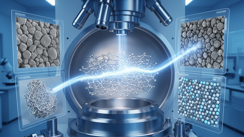
For most of the history of materials science, researchers have studied their specimens the way paleontologists study fossils: carefully, destructively, and always after the fact. A metal sample was deformed, fractured, heated, or corroded, and only then was it cut open and examined. The initial and final states could be compared, but everything that happened in between—the fleeting moments when dislocations moved, phases nucleated, or cracks began to form—was lost forever. A comprehensive review published in the Journal of Materials Science by Fei Liu of the State Key Laboratory of Nonlinear Mechanics at the Chinese Academy of Sciences and colleagues argues that in-situ characterization techniques have fundamentally broken through this static limitation, making it possible to watch the dynamic evolution of microstructures as it actually happens, even while a material is simultaneously subjected to multiple coupled fields such as stress, temperature, electric or magnetic fields, and reactive environments.
The review systematically surveys five major families of in-situ techniques, each occupying a different rung on the ladder of spatial resolution. At the top of that ladder, in terms of both scale and penetrating power, sit in-situ X-ray diffraction and synchrotron radiation methods, together with X-ray computed tomography. Diffraction probes the lattice itself, tracking changes in crystal structure, lattice parameters, and phase fractions with sub-nanometer precision. Synchrotron sources, which generate X-rays orders of magnitude brighter than laboratory tubes, allow these measurements to be collected in milliseconds or faster, so researchers can follow phase transformation kinetics as a material deforms. Computed tomography extends the view into three dimensions, reconstructing the internal architecture of a bulk component non-destructively and enabling so-called 4D studies in which the third spatial dimension is joined by time as the fourth.
The authors highlight striking examples of what these synchrotron methods can now reveal. In load-partitioning studies of multiphase alloys, in-situ diffraction can measure how each crystallographic phase in a composite or duplex material individually carries the applied stress, showing how the load is transferred between phases as deformation proceeds. In damage evolution studies of carbon fiber-reinforced silicon carbide composites, synchrotron in-situ tomography combined with deep learning has tracked coupled damage across a full oxygen concentration gradient at elevated temperature, even uncovering an oxygen-diffusion shielding mechanism that governs the thermo-mechanical-oxidative behavior of these ceramics. Similar approaches have mapped microcrack evolution in fiber-reinforced recycled concrete, characterized chloride-contaminated mortar under compression, and followed corrosion of aluminum alloys under combined thermal and chloride conditions.
Between the atomic and the component scales sits digital image correlation, or DIC, a technique that the review treats as the workhorse of full-field surface strain measurement. DIC works by comparing successive digital images of a specimen surface, typically decorated with a fine speckle pattern, and computing the displacement and strain fields that best map one image onto the next. The result is a complete two-dimensional or three-dimensional map of deformation, rather than a handful of point measurements. When performed through the lens of a scanning electron microscope, SEM-DIC can resolve strain fields down to the microstructural level, revealing how individual grains deform, where strain localizes, and where damage nucleates. The cited literature shows the breadth of this approach: measuring deformation kinematics during orthogonal cutting of titanium alloys, quantifying heterogeneous plastic deformation and so-called HDI strengthening in dual-phase steels, tracking strain localisation in particle-reinforced aluminum composites at high temperature, and even characterizing the mechanics of soft biological tissues such as the tricuspid valve.
The review then turns to electron microscopy-based methods, beginning with in-situ scanning electron microscopy itself. In an SEM, a tensile, fatigue, heating, or corrosion stage can be mounted inside the vacuum chamber, and the beam scans across the sample surface in real time. This allows researchers to watch crack initiation, propagation, and closure with sub-micrometer resolution while simultaneously correlating what they see with the applied load. The cited studies span fatigue short-crack growth in microscale single-crystal copper, slip-controlled fatigue crack growth in nickel-based bicrystal superalloys, hydrogen-charging effects on low-cycle fatigue of titanium alloys, and phase transformation and twinning mechanisms in interstitial high-entropy alloys. A growing trend noted in the review is automation and machine learning: spatiotemporal SEM image sequences are now being used to predict remaining fatigue life through domain-adaptive deep learning models, converting qualitative observation into quantitative forecasting.
Electron backscatter diffraction, EBSD, adds crystallographic information to the microscopic picture. Mounted on an SEM, an EBSD detector captures diffraction patterns from each scanned point on the surface and determines the local crystal orientation. In-situ EBSD experiments—deforming a sample inside the microscope and periodically mapping the orientation field—reveal how grains rotate, how deformation twins nucleate and grow, and how strain partitions among grains with different orientations. The review cites work on twinning in magnesium alloys, deformation mechanisms in high-entropy alloys, abnormal grain growth in nickel-based superalloys and high-purity cobalt, and the reverse transformation of austenite from lath martensite in steels. Quasi-in-situ EBSD, in which the sample is unloaded and remounted between mapping steps, extends these capabilities to fatigue and weld studies where continuous observation is impractical, and increasingly EBSD data are being fed directly into crystal plasticity finite element models to validate simulated grain reorientation trajectories.
At the finest scale lies in-situ transmission electron microscopy. TEM pushes spatial resolution to the atomic level, and modern instruments can be equipped with heating, cooling, tensile, nanoindentation, electrochemical, and irradiation stages. This means researchers can watch individual dislocations glide, stacking faults form, precipitates coarsen, grain boundaries migrate, and radiation damage accumulate—all in real time. The cited literature is remarkable in its scope: dislocation dynamics in high-entropy alloys analyzed with deep learning pipelines, irradiation-induced void swelling in nickel analyzed with computer vision, heavy-ion irradiation of zirconium alloys revealing interactions between defects and hydrogen atoms, lithium and sodium storage mechanisms in battery electrode materials observed during charging, and the growth of reaction layers in thin-film diffusion couples. Liquid-phase and cryo-TEM extend this capability to solutions and frozen states, enabling direct visualization of electroplating, ice nucleation, and phase separation.
What emerges from the review is not a picture of competing techniques but of complementary ones, each with defined applicable boundaries. X-ray methods penetrate bulk materials but average over larger volumes; electron microscopy resolves fine detail but only thin specimens or surfaces. DIC measures full strain fields but requires line-of-sight access to a prepared surface. The most powerful modern studies combine several techniques simultaneously—diffraction with DIC, SEM with EBSD, CT with digital volume correlation—to span the scales from lattice to component. The review emphasizes that this multiscale, real-time observational capability is what allows researchers to attack the central scientific problems of materials mechanics: load partitioning in multiphase alloys, three-dimensional damage evolution, phase transformation kinetics, and fatigue fracture, all under coupled multi-field service conditions that no static measurement can reproduce.
Yet the review is candid about four core challenges. The first is the inherent trade-off between spatial and temporal resolution: fast, high-resolution measurements demand brilliant sources and sensitive detectors, and improving one dimension often sacrifices the other. The second is the difficulty of reproducing extreme service environments—combining high temperature, reactive atmospheres, mechanical loading, and radiation in a single experimental window remains an engineering feat, and the sample environments themselves can introduce artifacts, as studies of electrochemical dead zones in in-situ XRD cells demonstrate. The third is the data deluge: in-situ experiments generate massive multimodal datasets—terabytes of images, diffraction patterns, and tomographic reconstructions—whose analysis increasingly depends on machine learning, from deep-learning segmentation of CT damage to automated dislocation tracking in TEM videos. The fourth is the non-uniqueness dilemma in model inversion: extracting microscopic parameters such as cohesive-zone laws from diffraction strain fields involves solving ill-posed inverse problems where many parameter sets can fit the same data.
The significance of this synthesis extends well beyond instrumentation. By establishing a critical theoretical foundation for dynamic, cross-scale observation, the review aims to support the construction of the processing–structure–property relationships that underpin modern materials design, from aerospace superalloys and fusion reactor materials to batteries, cement, and additive-manufactured components. When engineers can see, in real time and across six orders of magnitude in length scale, how a microstructure actually evolves under the coupled fields of real service, they can design materials from mechanism rather than from correlation. In that sense, the era of watching materials think—deform, transform, and fail in real time—is not merely a technical achievement but a conceptual shift in what it means to understand a material.
Subject of Research: Cross-scale in-situ characterization techniques for tracking dynamic microstructural evolution in materials under coupled multi-field conditions
Article Title: Review:cross-scale in-situ characterization techniques for dynamic evolution of microstructures in materials under multi-field coupling
Article References: Liu, F., Zhang, M., Zhang, F., Li, J., Xie, H., Wang, Q., & Huan, Y. (2026). Review:cross-scale in-situ characterization techniques for dynamic evolution of microstructures in materials under multi-field coupling. Journal of Materials Science, 61(41), 31707-31770. https://doi.org/10.1007/s10853-026-13674-z
Image Credits: AI Generated
DOI: 10.1007/s10853-026-13674-z
Keywords: in-situ characterization, synchrotron X-ray diffraction, X-ray computed tomography, digital image correlation, scanning electron microscopy, electron backscatter diffraction, transmission electron microscopy, microstructure evolution, multi-field coupling, phase transformation, fatigue and damage, materials characterization
Cite Scienmag News
Denise Maddox. (October 19, 2026). Watching Materials Transform in Real Time: A Review of Cross-Scale In-Situ Characterization. Scienmag. https://scienmag.com/watching-materials-transform-in-real-time-a-review-of-cross-scale-in-situ-characterization/
Denise Maddox. "Watching Materials Transform in Real Time: A Review of Cross-Scale In-Situ Characterization." Scienmag, 19 October 2026, https://scienmag.com/watching-materials-transform-in-real-time-a-review-of-cross-scale-in-situ-characterization/. Accessed 19 October 2026.
Denise Maddox. "Watching Materials Transform in Real Time: A Review of Cross-Scale In-Situ Characterization." Scienmag. October 19, 2026. https://scienmag.com/watching-materials-transform-in-real-time-a-review-of-cross-scale-in-situ-characterization/


