


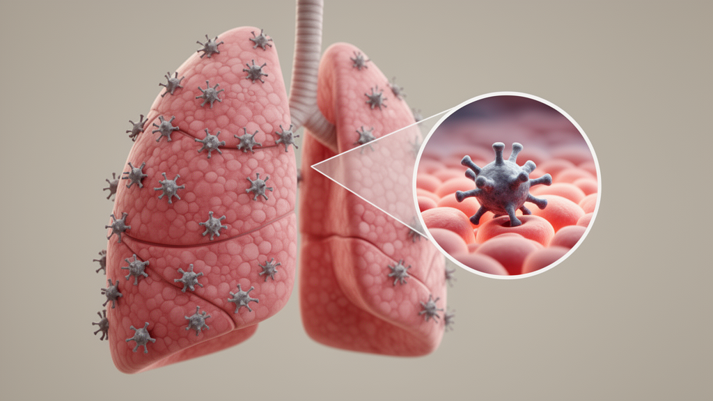
More than two years after the acute phase of COVID-19 has resolved, some patients continue to struggle with breathlessness, cough, and abnormal chest scans that stubbornly refuse to clear. What is actually happening inside the lungs of these people has remained largely a mystery, because direct sampling of the deep lung is invasive and rarely performed outside of specialized centers. A new study from investigators at Indiana University, published in Frontiers in Microbiology, offers one of the most detailed looks yet at the inflammatory environment of the alveoli in patients with suspected post-COVID-19 lung disease, and its findings point to a disease that is far more heterogeneous than its clinical presentation suggests.
The research team, led by pulmonologist Homer L. Twigg III, took advantage of a unique resource: a CLIA-approved clinical bronchoalveolar lavage laboratory that serves clinicians across central Indiana. Bronchoalveolar lavage, or BAL, involves threading a bronchoscope into the airways and instilling saline to wash out cells and soluble proteins from the alveolar spaces, the site where gas exchange occurs. Since October 2020, the laboratory had been receiving BAL fluid from patients undergoing bronchoscopy as part of their clinical work-up for persistent pulmonary symptoms after COVID-19. The only requirement for inclusion in the study cohort was a documented positive SARS-CoV-2 PCR test at some point before the lavage was performed.
By May 2022, the investigators had collected 37 BAL samples from 37 different patients. Five of these were obtained less than 28 days after a positive test and were classified as acute infections, while the remaining 32 were collected between 28 and 730 days after diagnosis, with a median of 178 days. The samples were processed by cytocentrifugation to generate cell differentials, and specimens contaminated with more than 15 percent buccal mucosal or airway epithelial cells were excluded. Two control groups were used for comparison: five healthy volunteers undergoing research bronchoscopy, and seven patients whose clinical BAL samples predated the pandemic entirely.
The cellular analysis revealed that suspected post-COVID-19 lung disease splits into three distinct inflammatory phenotypes. The first was essentially a normal BAL cell differential, with no increase in lymphocytes and minimal neutrophil accumulation. The second was a neutrophilic predominant pattern, defined by neutrophils making up at least 10 percent of the total nucleated cell population and exceeding the lymphocyte fraction. The third was a lymphocytic predominant pattern, in which lymphocytes accounted for at least 15 percent of cells and outnumbered neutrophils. Among the 12 patients with elevated lymphocytes, 8 had markedly inverted CD4-to-CD8 ratios, a finding classically associated with hypersensitivity pneumonitis, nonspecific interstitial pneumonia, chronic viral infections, and cryptogenic organizing pneumonia. The two patients with elevated rather than inverted ratios had underlying conditions, sarcoidosis in one case and mycobacterial antigen-associated hypersensitivity pneumonitis in the other, both known to drive CD4-predominant alveolitis.
Perhaps the most striking result came from the search for viral remnants. Using a commercial ELISA with a sensitivity of 0.256 picograms per milliliter, the team measured SARS-CoV-2 spike protein in acellular BAL fluid. Spike protein was readily detected in unconcentrated lavage fluid from the post-COVID patients compared with controls, and the difference remained significant even after correcting for dilution by expressing concentrations as picograms per milliliter of epithelial lining fluid, calculated using the urea method developed by Rennard and colleagues. Remarkably, spike protein was detected up to 627 days after the initial diagnostic test, supporting the hypothesis that residual viral proteins persist in the lung long after acute infection has cleared. The authors note that expanded data on persistent spike and nucleocapsid proteins, as well as viral nucleic acids, in this same cohort are described in a separate submitted manuscript.
To probe the pulmonary antibody response, the investigators turned to the PepSeq platform, a highly multiplexed serological technique that uses libraries of thousands of DNA-barcoded peptides with next-generation sequencing readout. For these experiments, a human virome-wide assay comprising 15,000 peptides was employed, with samples sequenced to a depth of more than 50 reads per peptide and replicate samples showing a median Z-score concordance of 0.94. Of the first 23 post-COVID subjects tested, 14, or 61 percent, had anti-SARS-CoV-2 antibodies in their BAL fluid, including many with polyclonal responses targeting epitopes on both spike and nucleocapsid proteins. Because the unstructured 30-mer peptides used in PepSeq cannot generally detect antibodies against conformational epitopes, the team also measured neutralizing activity with the cPass assay, which tests the ability of a sample to block binding between spike protein and the ACE2 receptor. Eleven of the 37 BAL samples, or 30 percent, exceeded the assay’s 30 percent inhibition cutoff, while no control sample exceeded 18 percent, and there was complete concordance between neutralizing antibody detection and PepSeq positivity.
The clinical correlations, however, proved more elusive. All subjects underwent chest CT scanning before bronchoscopy, and the scans were reviewed blindly by a NIOSH B Reader radiologist and grouped into predominantly ground glass opacities, predominantly fibrotic or interstitial changes, an equal mixture of both, or other patterns. No clear association emerged between radiographic appearance and the type of inflammatory milieu found in the lung. Long-term outcomes, tracked through chart review as of January 1, 2026, included vital status, need for lung transplant, and need for supplemental oxygen. The BAL inflammatory pattern was unable to predict long-term outcome in this underpowered analysis, although an intriguing signal appeared: no early adverse outcomes were observed among subjects with a normal BAL differential. In unadjusted logistic regression, the odds of a good outcome relative to the neutrophilic pattern were 2.13 for the normal group and 2.24 for the lymphocytic group, with confidence intervals that crossed one.
The findings carry significant implications for how post-COVID-19 lung disease is studied and treated. Because the NASEM definition of long COVID adopted by the NIH RECOVER initiative is purely clinical, relying on symptoms and radiographic findings after documented infection, patients grouped together under that umbrella may in fact harbor fundamentally different processes. The authors suggest that patients with a CD8-predominant lymphocytic pattern may have cryptogenic organizing pneumonia and could respond to corticosteroids, while those with neutrophilic inflammation may have the usual interstitial pneumonia pattern described in surgical lung biopsies of post-COVID patients. Patients with normal lavage fluid, who underwent bronchoscopy because of persistent CT abnormalities, likely have stable scar tissue from their acute infection, which could explain why some post-COVID fibrotic changes fail to progress and may even resolve, echoing the experience after SARS-CoV-1.
The persistence of spike protein also fits into a broader picture of chronic viral antigen stimulation. Prior work has documented residual viral proteins long after other infections, including HIV Nef protein in the lungs of aviremic patients and residual antigen presentation following influenza. Autopsy studies performed a median of 39 days after infection found persistent viral genomic RNA in nearly all patients, and a systematic review detected SARS-CoV-2 RNA in various tissues in 5 to 59 percent of subjects up to six months after acute infection. Persistent antigen can sustain chronic inflammation through multiple mechanisms, including ongoing B and T cell responses, T cell exhaustion and immunosenescence, direct stimulation of macrophages through TLR2, and activation of the complement system, which could plausibly drive the neutrophilic inflammation seen in a third of the patients. Notably, some form of immunosuppression was prevalent in the cohort, affecting 28 of 37 subjects, which the authors suggest may contribute to failure to clear the virus.
The investigators are careful to acknowledge the limitations of their work. The cohort is small and underpowered for linking inflammatory patterns to outcomes, the samples were collected relatively early in the pandemic before widespread vaccination and when more virulent strains circulated, and the cPass 30 percent cutoff was developed for plasma rather than BAL fluid, making the neutralization findings exploratory. There is also no comparison cohort of previously infected patients without chronic lung disease. Still, the study demonstrates that post-COVID-19 lung disease exists in several immunologically distinct forms, that viral proteins can linger in the lung for up to two years, and that clinical presentation alone may be an unreliable guide to the biology underneath. Whether residual SARS-CoV-2 components drive these phenotypes, and whether SARS-CoV-2-specific immune responses can ameliorate the disease, is currently under study.
Subject of Research: Inflammatory and immunologic profiling of bronchoalveolar lavage fluid in patients with post-COVID-19 lung disease
Article Title: Inflammatory and immunologic analysis of bronchoalveolar lavage from patients with post-COVID-19 lung disease
Article References: Twigg, H. L., III, Wilson, R., Yu, A. Q., Li, W., Altin, J. A., Tarver, R. D., Knight, I., Smith, P. A., Fraser, M. E., Castelluccio, P., & Rie, J. (2026). Inflammatory and immunologic analysis of bronchoalveolar lavage from patients with post-COVID-19 lung disease. Frontiers in Microbiology, 17, Article 1923299. https://doi.org/10.3389/fmicb.2026.1923299
Image Credits: AI Generated
DOI: 10.3389/fmicb.2026.1923299
Keywords: long COVID, post-COVID-19 lung disease, bronchoalveolar lavage, SARS-CoV-2, spike protein, viral persistence, neutralizing antibodies, PepSeq, pulmonary inflammation, interstitial lung disease, alveolar macrophages, immunosenescence
Cite Scienmag News
Kristina Jarvis. (October 19, 2026). Lingering Viral Spike Protein Found in the Lungs of Long COVID Patients. Scienmag. https://scienmag.com/lingering-viral-spike-protein-found-in-the-lungs-of-long-covid-patients/
Kristina Jarvis. "Lingering Viral Spike Protein Found in the Lungs of Long COVID Patients." Scienmag, 19 October 2026, https://scienmag.com/lingering-viral-spike-protein-found-in-the-lungs-of-long-covid-patients/. Accessed 19 October 2026.
Kristina Jarvis. "Lingering Viral Spike Protein Found in the Lungs of Long COVID Patients." Scienmag. October 19, 2026. https://scienmag.com/lingering-viral-spike-protein-found-in-the-lungs-of-long-covid-patients/


