


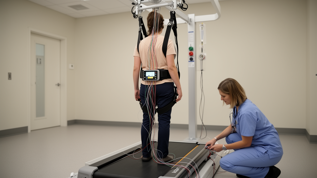
For people living with spinal cord injury, one of the most striking developments in modern neurorehabilitation is epidural spinal cord stimulation, a technique in which electrodes implanted over the dorsal surface of the spinal cord deliver electrical pulses that can re-enable standing, walking, arm control, and even regulation of blood pressure and bladder function. A new narrative review published in Frontiers in Neurology by researchers from Henan Medical University, Zhengzhou Central Hospital Affiliated to Zhengzhou University, and Xinjiang Medical University takes a hard look at how the technique actually works — and, more importantly, how the choice of stimulation parameters determines what it can achieve. The verdict is both exciting and sobering: the technology works, but the field’s evidence base remains fragmented, dominated by small uncontrolled studies, and far from delivering a universal prescription.
The biological logic behind the approach is elegant. A spinal cord injury does not necessarily destroy the networks below the lesion; it disconnects them from the brain. Locomotor circuits, including central pattern generators capable of producing rhythmic stepping, often remain anatomically intact but functionally dormant. Epidural stimulation recruits large-diameter sensory afferents in the dorsal roots and dorsal columns, raising the excitability of spinal motor pools so that weak residual descending commands can be converted into functional movement — even in people classified as motor-complete. Crucially, recent work combining human electromyography with primate and computational data shows that stimulation pulses and voluntary effort interact multiplicatively rather than additively: a pulse train that is subthreshold on its own becomes effective only when residual descending input depolarizes motoneurons into a permissive range. This defines a restricted, individual “functional parameter space” that narrows as lesion severity increases — which is why no single parameter set can work for everyone.
The review maps six parameter domains onto clinical outcomes. Electrode placement is the most consequential: lumbosacral paddle arrays at the T11–L1 vertebral level, overlying the L1–S1 spinal segments, are the standard for standing, walking, and pelvic-organ function, while cervical stimulation targeting the C4–T2 enlargement is required for arm and hand recovery. Frequency shows a clear functional segregation: low frequencies of roughly 15–60 Hz facilitate motor output, whereas kilohertz-range stimulation — around 1.2 kHz — acts as a reversible conduction block that suppresses spasticity. Intensity is titrated against physiological anchors such as motor threshold or the slope ratio of muscle recruitment curves, and pulse widths typically fall between 210 and 500 microseconds, though the review notes that pulse width is frequently omitted from landmark publications, making dose comparisons across studies nearly impossible.
Two distinct parameter philosophies have emerged for restoring movement. The first is tonic, individually titrated lumbosacral stimulation embedded in months of intensive activity-based training — the approach behind the field’s most famous results, including full weight-bearing standing in motor-complete paraplegia and volitional leg movement in all thirteen participants of one landmark series. The second is programmed, biomimetic stimulation: spatiotemporal programs that sequence contacts in time with the gait cycle, dual-mode protocols that combine kilohertz spasticity control with low-frequency facilitation, and closed-loop systems that adjust stimulation in real time. Spatiotemporal approaches enabled adaptive walking within a week in some motor-incomplete participants and restored walking capacity in people with complete sensorimotor paralysis, while a fully implanted brain–spine interface that decodes cortical motor intent has restored natural walking and stair climbing, with calibration stable over more than a year of home use.
One of the review’s most interesting tensions concerns proprioception. Computational, animal, and human evidence shows that continuous stimulation can transiently block proprioceptive input — a kind of deafferentation that abolishes leg-position perception — which motivated the shift toward burst and spatiotemporal profiles that preserve sensory feedback. Yet the tonic, continuous approach produced the field’s most-cited functional gains in motor-complete patients. The authors reconcile this by noting that the proprioception-blocking demonstrations largely involved motor-incomplete participants with preserved sensory channels worth protecting, whereas in complete injuries there may be little proprioceptive transmission left to disrupt. The trade-off, they conclude, is real but task- and individual-dependent, not absolute.
Autonomic applications are quietly becoming the most mature part of the field. Blood-pressure management now has the most standardized parameter set of any indication: a lower-thoracic “hemodynamic hotspot” at the last three thoracic segments, 120 Hz stimulation, roughly 300-microsecond pulses, and amplitudes titrated to the pressor response under continuous or closed-loop control. This configuration — which head-to-head testing showed outperforms conventional lumbosacral targeting — produced durable hemodynamic stability and allowed discontinuation of pressor medications in a fourteen-participant series. Bladder outcomes are consistent but confined largely to a single center, with around 30 Hz identified as optimal for voiding efficiency, while bowel benefits remain secondary, self-reported endpoints without a dedicated parameter set.
Upper-limb recovery, spasticity, and pain tell a more cautionary tale. Cervical epidural stimulation is mechanistically strong — primate work shows selective recruitment of upper-limb motoneurons via sensory afferents — but the human evidence rests on a single two-participant preprint, while the only large pivotal trial in the arm-and-hand space used non-invasive transcutaneous stimulation and led to the field’s first regulatory authorization. For spasticity, the historical 100–130 Hz tonic paradigm and the newer kilohertz-block dual-mode strategy coexist without any dose-finding trial to adjudicate between them. And for neuropathic pain after spinal cord injury, the review finds no SCI-specific parameter evidence at all: paradigms are borrowed directly from chronic-pain practice, a systematic review graded the entire evidence base as low quality with no randomized trials, and the only modern case series reported satisfactory relief in just two of eight patients.
The methodological weaknesses of the literature loom large. A scoping review of 103 human studies found only one randomized controlled trial, with 70 percent of studies enrolling ten or fewer participants and stimulation parameters incompletely described across many reports. Every included epidural publication was supportive of the intervention, with no reports of significant failures — a pattern the authors flag as strongly suggestive of publication bias. Meanwhile, the field’s most rigorous test to date, a triple-blind sham-controlled trial of transcutaneous stimulation plus locomotor training in fifty participants, found no added benefit over training alone, though the authors stress this constrains only the non-invasive modality and cannot be extrapolated to epidural protocols, which offer far greater spatial selectivity. Amplitudes reported in volts and milliamperes are not directly comparable, further blocking quantitative synthesis.
The review is also refreshingly clear about what stimulation-plus-training means. Virtually every positive study delivers stimulation embedded in intensive, task-specific rehabilitation — from roughly 80 to as many as 278 training sessions — and the authors argue this coupling is inherent to the therapy, not a confound. Stimulation changes the excitability state of spinal networks, but training supplies the task-specific information needed to organize those circuits for function. Gains are strikingly task-specific: robotic stand training improved standing without transferring to sitting control, while trunk-specific stimulation programmed for sitting improved sitting stability. Reported “optimal parameters,” the authors suggest, may index residual anatomy as much as any universal dose–response, arguing for imaging- and electrophysiology-based patient stratification.
Where does this leave the field? The authors’ prescription is unambiguous: adequately powered, sham-controlled randomized trials of epidural stimulation; a mandatory, uniform parameter-reporting checklist covering electrode configuration, frequency, amplitude with units, pulse width, waveform, duty cycle, and concurrent training dose; routine imaging-based stratification; prospective long-term safety registries; and explicit cost-effectiveness analysis. Closed-loop, intent-driven systems — brain–spine interfaces, bidirectional perilesional stimulation with deep-learning-optimized parameters, and AI-assisted titration — represent the clearest technical trajectory, recasting stimulation parameters as dynamic, intent-contingent variables rather than fixed clinician-programmed constants. Until controlled trials catch up, the review concludes, epidural stimulation remains best understood as an enabling component of a combined stimulation-plus-training program: a technology capable of remarkable individual recoveries, still awaiting the evidence architecture that will turn those anecdotes into reproducible, evidence-graded medicine.
Subject of Research: Stimulation parameters of epidural spinal cord stimulation for functional recovery after spinal cord injury
Article Title: Stimulation parameters of epidural spinal cord stimulation for functional recovery after spinal cord injury: a narrative review
Article References: Wang, J., Bieshanbieke, Y., Wang, H., Yang, J., Ji, S., Li, Y., Gu, H., & Lou, Y. (2026). Stimulation parameters of epidural spinal cord stimulation for functional recovery after spinal cord injury: a narrative review. Frontiers in Neurology, 17, Article 1952514. https://doi.org/10.3389/fneur.2026.1952514
Image Credits: AI Generated
DOI: 10.3389/fneur.2026.1952514
Keywords: epidural spinal cord stimulation, spinal cord injury, neuromodulation, functional recovery, stimulation parameters, neurorehabilitation, spatiotemporal stimulation, brain-spine interface, autonomic function, spasticity, locomotion, closed-loop stimulation
Cite Scienmag News
Cassandra Pierce. (October 19, 2026). Electrical Stimulation of the Spine Can Restore Movement After Paralysis — But the Recipe Is Still Being Written. Scienmag. https://scienmag.com/electrical-stimulation-of-the-spine-can-restore-movement-after-paralysis-but-the-recipe-is-still-being-written/
Cassandra Pierce. "Electrical Stimulation of the Spine Can Restore Movement After Paralysis — But the Recipe Is Still Being Written." Scienmag, 19 October 2026, https://scienmag.com/electrical-stimulation-of-the-spine-can-restore-movement-after-paralysis-but-the-recipe-is-still-being-written/. Accessed 19 October 2026.
Cassandra Pierce. "Electrical Stimulation of the Spine Can Restore Movement After Paralysis — But the Recipe Is Still Being Written." Scienmag. October 19, 2026. https://scienmag.com/electrical-stimulation-of-the-spine-can-restore-movement-after-paralysis-but-the-recipe-is-still-being-written/


