


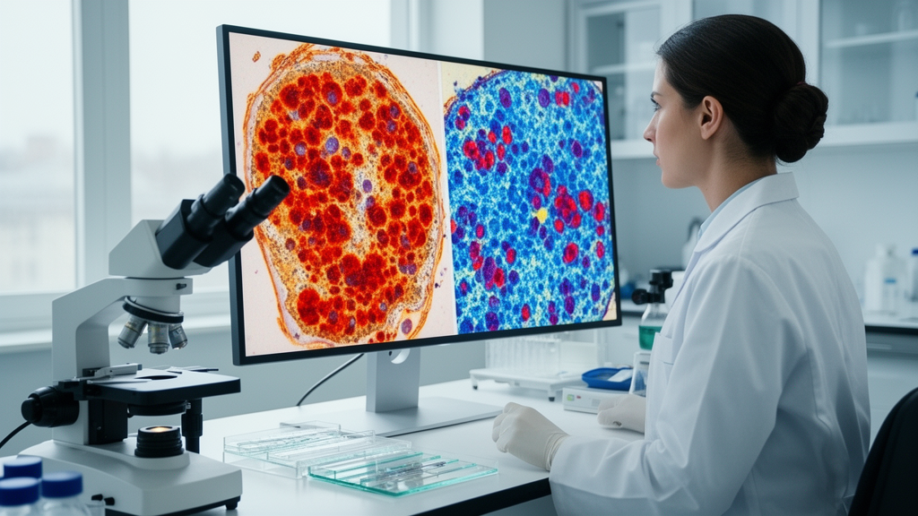
Habitat imaging analysis represents a departure from the conventional radiomics paradigm, in which a single set of features is extracted from a manually or automatically delineated tumour volume and treated as representative of the whole lesion. Instead, habitat approaches recognise that tumours are spatially heterogeneous structures composed of biologically distinct subregions. By applying clustering algorithms to voxel-level imaging data, these methods partition a tumour into a small number of intratumoural habitats, each characterised by a shared pattern of imaging attributes such as attenuation on computed tomography, signal intensity on magnetic resonance imaging, or tracer uptake on positron emission tomography. The underlying premise is that these subregions correspond, at least approximately, to underlying biological differences within the tumour, including variations in cellularity, necrosis, hypoxia, vascularity, and proliferative activity. In head and neck cancer, where anatomical complexity and tissue-to-tissue contrast issues complicate imaging interpretation, this spatially aware framework has attracted growing interest.
The systematic review by Ai and colleagues provides a timely synthesis of this rapidly evolving literature. By searching PubMed, Web of Science, and Scopus from database inception through 30 January 2026, the authors captured twenty-five original studies published between 2019 and 2026, a period that encompasses essentially the entire history of habitat analysis applied to head and neck tumours. This short time frame is itself informative: the field is young, and the pace of publication suggests accelerating interest. Yet the review also reveals important structural weaknesses in the evidence base. Only ten of the twenty-five studies included external validation datasets, meaning that the majority of reported models were evaluated solely on data drawn from the same institutions or patient cohorts used to develop them. This is a well-recognised threat to the generalisability of imaging prediction models, because internally validated performance frequently overstates what can be expected when a model is applied to patients from different scanners, protocols, or populations.
The dominance of nasopharyngeal carcinoma among the reviewed studies, with nine of the twenty-five articles devoted to it, reflects both the epidemiology of head and neck cancer globally and the research priorities of the regions where habitat imaging has been most actively pursued. Nasopharyngeal carcinoma is endemic in parts of southern China and Southeast Asia, and its deep anatomical location makes biopsy-based assessment of intratumoural heterogeneity challenging, which in turn creates a strong incentive for non-invasive imaging surrogates. Computed tomography was the most frequently used modality, appearing in nine studies, which is notable given that magnetic resonance imaging is often considered the mainstay of head and neck tumour assessment. The popularity of computed tomography in habitat work likely relates to its quantitative attenuation values, which lend themselves naturally to voxel-wise clustering, and to its widespread availability in routine staging workflows, making retrospective data collection straightforward.
Methodologically, K-means clustering was the predominant habitat technique, employed in twenty of the twenty-five studies. K-means is computationally simple, easy to implement, and produces readily interpretable partitions, which explains its popularity. However, its ubiquity also raises questions about whether the field has explored the full methodological space. Alternative clustering approaches, including Gaussian mixture models, fuzzy c-means, and hierarchical methods, may capture different aspects of tumour heterogeneity, and the optimal number of habitats to extract remains largely a matter of empirical choice rather than biological principle. The review’s observation that most studies selected a small number of subregions, typically two to four, is consistent with the practical need to balance model complexity against the risk of overfitting, but it also means that finer-grained biological variation may be lost in the coarser partitions that most analyses employ.
The finding that habitat imaging analysis performed consistently well, with areas under the curve exceeding 0.8, in predicting pathological and immunohistochemical biomarkers, tumour characterisation, and nodal status is encouraging. These are clinically meaningful tasks. Pathological and immunohistochemical markers, such as measures of proliferation, hypoxia, or immune infiltration, currently require tissue sampling, and non-invasive surrogates could in principle reduce the need for biopsy or guide more targeted sampling. Nodal status assessment is central to staging and treatment planning in head and neck cancer, and any imaging tool that improves on conventional size-based criteria could have real clinical impact. The fact that habitat-derived features outperformed conventional radiomics or clinical models in these domains suggests that explicitly modelling intratumoural spatial heterogeneity captures information that whole-tumour averaging discards.
In contrast, the variable results for treatment response prediction warrant careful interpretation. Predicting how an individual tumour will respond to radiotherapy, chemotherapy, or combined modalities is arguably the most valuable application imaginable for imaging-based heterogeneity analysis, because treatment de-escalation and escalation decisions in head and neck cancer carry significant trade-offs between tumour control and toxicity. The inconsistency of habitat models in this domain may reflect genuine biological complexity: treatment response is influenced by tumour factors, host factors, and treatment factors in ways that a static pretreatment imaging snapshot may not fully capture. It may also reflect methodological issues, including small sample sizes, heterogeneous endpoints such as pathological complete response versus progression-free survival, and differing time points at which imaging was performed relative to therapy.
The observation that combining multiple models only slightly improved performance in most studies is a sobering counterpoint to the prevailing enthusiasm for multimodal and multi-model integration. In many areas of imaging research, combining complementary data sources is assumed to yield additive or even synergistic gains. The review’s finding raises a practical question: if the incremental benefit of integrating habitat models with conventional radiomics or clinical predictors is marginal, does the added complexity, computational cost, and validation burden justify clinical adoption? This question is particularly pertinent in head and neck cancer, where clinical decision-making already integrates a rich array of clinical, pathological, and imaging information, and where any new tool must demonstrate clear added value over existing standards.
The heterogeneity of the current evidence base, which the authors explicitly highlight, deserves emphasis. The twenty-five included studies varied in tumour origin, imaging modality, clustering features, habitat methods, number of subregions, and clinical applications. Such heterogeneity makes it difficult to draw firm conclusions about which habitat approaches work best, for which tumour types, and for which clinical questions. It also complicates meta-analysis, since pooling results across studies with fundamentally different designs risks producing misleading summary estimates. The Radiomics Quality Score, referenced in the review’s abbreviations, is one tool that has been developed to assess the methodological rigour of imaging studies, and its application in this context underscores the field’s growing awareness of the need for standardisation.
Looking forward, the review’s call for prospectively conducted and externally validated studies is both appropriate and necessary. Prospective designs would allow standardised imaging protocols, predefined endpoints, and sample sizes powered to detect clinically meaningful effects, all of which are difficult to achieve retrospectively. External validation across multiple institutions, ideally with diverse patient populations and scanner types, would establish whether the promising internal performance of habitat models translates to real-world settings. Standardisation of habitat methodology, including consensus on clustering algorithms, feature selection, and subregion numbering, would further strengthen the evidence base and facilitate meaningful comparison across studies.
The open-access publication of this review under a Creative Commons Attribution licence ensures that its findings are freely available to clinicians, researchers, and patients worldwide, which is particularly valuable for a field in its infancy where broad access to methodological critiques can shape research priorities. The involvement of authors from both the Chinese University of Hong Kong and the University of Hong Kong reflects the regional concentration of head and neck cancer expertise and imaging research capacity, and their collaboration across radiology and oral and maxillofacial radiology disciplines illustrates the multidisciplinary nature of this work. As habitat imaging analysis matures, the transition from retrospective proof-of-concept studies to prospective, multicentre validation trials will determine whether the consistent performance observed in biomarker prediction and nodal assessment can be harnessed to improve outcomes for patients with head and neck cancer, and whether the more uncertain domain of treatment response prediction can be refined to the point where it informs individualised therapeutic decisions.
Subject of Research: Habitat imaging analysis for head and neck cancer
Article Title: Current evidence in application of habitat imaging analysis in head and neck cancer: a systematic review
Article References: Ai, Q. Y. H., Cai, Z., Leung, H. S., Kwok, H. M., So, T. Y., & Hung, K. F. (2026). Current evidence in application of habitat imaging analysis in head and neck cancer: a systematic review. BMC Medical Imaging. https://doi.org/10.1186/s12880-026-02777-3
Image Credits: AI Generated
DOI: 10.1186/s12880-026-02777-3
Keywords: habitat imaging, head and neck cancer, radiomics, nasopharyngeal carcinoma, machine learning, CT imaging, MRI, PET, K-means clustering, systematic review, tumour heterogeneity, biomarker prediction
Cite Scienmag News
Nathaniel Bowman. (October 18, 2026). Habitat Imaging Analysis Shows Promise for Head and Neck Cancer Care, Systematic Review Finds. Scienmag. https://scienmag.com/habitat-imaging-analysis-shows-promise-for-head-and-neck-cancer-care-systematic-review-finds/
Nathaniel Bowman. "Habitat Imaging Analysis Shows Promise for Head and Neck Cancer Care, Systematic Review Finds." Scienmag, 18 October 2026, https://scienmag.com/habitat-imaging-analysis-shows-promise-for-head-and-neck-cancer-care-systematic-review-finds/. Accessed 18 October 2026.
Nathaniel Bowman. "Habitat Imaging Analysis Shows Promise for Head and Neck Cancer Care, Systematic Review Finds." Scienmag. October 18, 2026. https://scienmag.com/habitat-imaging-analysis-shows-promise-for-head-and-neck-cancer-care-systematic-review-finds/


