


A blood clot that lodges in the arteries of the lungs can turn a routine recovery into a life-threatening emergency within minutes. Pulmonary embolism, the condition caused when clots that typically form in the deep veins of the legs break free and travel to the lungs, ranks as the third most common cardiovascular killer in the United States, behind only heart attack and stroke. Once a large clot blocks the pulmonary arteries, oxygen exchange is disrupted and the heart is suddenly forced to pump against a dangerous obstruction, a strain that can rapidly become fatal. Now, a team of engineers and vascular surgeons at the University of Michigan, funded by a $3.74 million grant from the National Institutes of Health, is developing a device designed to destroy such clots from the inside, using a form of sound waves that twist as they travel.
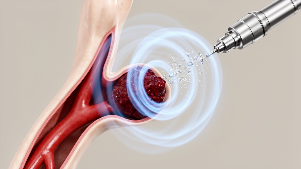
The technology, which the researchers call vortex ultrasound, centers on a tiny ultrasound source mounted at the tip of a catheter. In the intended procedure, a physician would thread the catheter through the patient’s blood vessels until the device reaches the clot itself. Once in position, the transducer would emit acoustic waves that rotate in a swirling, vortex-like pattern rather than propagating in the simple, linear fashion of conventional ultrasound. According to the research team, this twisting motion attacks the clot’s internal structure more effectively than standard acoustic energy, and the mechanism appears to depend in part on a well-known physical phenomenon: the rapid creation and collapse of bubbles within the clot material.
That bubble-driven process, known in acoustics as cavitation, is central to why the team believes swirling waves can dismantle clots so quickly. When intense ultrasound passes through a medium, microscopic bubbles can nucleate within it, and their oscillation and collapse generate intense localized mechanical forces. Inside a soft, fibrous clot, those forces can tear apart the protein mesh that holds the mass together, fragmenting it into pieces small enough to be handled by the body’s normal processes. The researchers suggest that the swirling character of the vortex waves accelerates this internal disruption, allowing the clot to break apart from within rather than being scraped or pulled away from the vessel wall.
Protecting that vessel wall is one of the device’s most important design goals. The pulmonary arteries are delicate structures, and one of the major drawbacks of existing catheter-based mechanical thrombectomy systems is the risk that a rigid removal tool could tear the vessel wall during extraction of the clot. By breaking the clot into fragments acoustically rather than grabbing and pulling it, the vortex ultrasound approach aims to minimize mechanical contact with the tissue around it. The team reports that early tests indicate the twisted waves can fragment clots without damaging the blood vessel walls, a property that would address one of the sharpest trade-offs in current emergency care.
The clinical context explains why such a device is urgently sought. Today’s standard options all involve significant compromises. Clot-dissolving drugs, known as thrombolytics, can take up to 24 hours to work and fail roughly a third of the time, and they carry a serious risk of internal bleeding because they act systemically on the patient’s clotting system rather than targeting the obstructing clot alone. Mechanical catheter devices trade the bleeding risk for a different hazard: the possibility of tearing fragile vessel walls during removal. In the most severe cases, surgeons must resort to open-chest surgery to extract the clot directly, an option reserved for the gravest presentations because of its invasive nature and associated risks.
External ultrasound might seem like an obvious middle ground, since applying sound waves from outside the body would avoid both the bleeding risks of drugs and the mechanical risks of catheters. In practice, however, noninvasive ultrasound struggles to deliver sufficient energy accurately to a target deep inside the body, and in the lungs it faces an additional obstacle: the organ’s internal air-filled structures scatter and interfere with ultrasound waves, rendering the approach largely ineffective there. The Michigan team’s insight is to change both variables at once. By altering the type of ultrasound employed and delivering it from inside the blood vessel, directly at the clot, the device sidesteps the propagation problem entirely while concentrating the acoustic energy where it is needed.
The speed of the approach is perhaps its most striking feature. In early proof-of-concept testing, the research team demonstrated that vortex ultrasound could restore blood flow through a three-inch-long blood clot in just eight minutes, using blood from cows in a mock circulatory setup. Preliminary results further suggest that the twisted ultrasound waves could break up clots in as little as 30 minutes in more clinically representative scenarios, a dramatic improvement over the many hours sometimes required for pharmacological thrombolysis. In pulmonary embolism, where the heart is under acute strain, that difference in time is not merely a matter of convenience; it could be the difference between recovery and catastrophic cardiovascular collapse.
The new five-year, $3.74 million NIH award will fund the work needed to move the technology from the laboratory bench toward the clinic. Over the course of the project, the team plans to refine the catheter design, optimize the shape and frequency of the ultrasound waves emitted by the miniature transducer, and rigorously evaluate both short- and long-term safety of the treatment in pigs, a standard large-animal model whose cardiovascular anatomy approximates that of humans closely enough to test interventional devices. Those safety studies will be critical, because any device intended to operate inside pulmonary arteries must demonstrate that it can destroy clots while leaving the surrounding tissue unharmed, both immediately after treatment and over extended follow-up periods.
The project brings together specialists whose expertise spans the full arc from acoustic physics to surgical practice. Principal investigator Chengzhi Shi, an associate professor of mechanical engineering at the University of Michigan, specializes in physical acoustics, metamaterials and ultrasound technologies for medical treatment, providing the core physics behind the swirling wave generation. Clinical guidance comes from Daniel Myers, the Conrad and Caroline Jobst Research Professor of Vascular Surgery, and Andrea Obi, the Wakefield Family Research Professor, both of the U-M Medical School, who help ground the engineering in the realities of treating patients with dangerous clots. The team also includes Zhen Xu, the Li Ka Shing Endowed Professor of Biomedical Engineering at U-M, whose work in therapeutic ultrasound adds depth to the acoustic design effort, and Xiaoning Jiang, professor of mechanical and aerospace engineering at North Carolina State University, a specialist in ultrasonic transducer technology.
For Shi, the motivation is straightforward: the goal is to treat clots in the lungs more quickly and safely, while potentially offering an alternative to open-chest surgery for patients with the most severe pulmonary embolisms. If the five-year program succeeds in demonstrating safety and effectiveness in animal models, vortex ultrasound could join a small but growing family of catheter-delivered, energy-based therapies that are reshaping emergency vascular medicine. For the tens of thousands of patients who develop pulmonary embolism each year, a treatment that can clear a deadly obstruction from the lungs in minutes, through a catheter rather than an open chest, would represent a fundamental shift in what is possible in the critical first hours of the disease.
Subject of Research: Vortex ultrasound catheter technology for treating pulmonary embolism
Article Title: Treating life-threatening blood clots in minutes with swirling sound waves
Article References: Treating life-threatening blood clots in minutes with swirling sound waves. (n.d.). Original publication
Image Credits: AI Generated
DOI: Not provided
Keywords: pulmonary embolism, vortex ultrasound, blood clots, catheter, thrombolysis, cavitation, University of Michigan, NIH grant, vascular surgery, mechanical engineering, therapeutic ultrasound, emergency medicine
Cite Scienmag News
Ophelia Keating. (October 18, 2026). Swirling Sound Waves Could Shatter Deadly Lung Clots in Minutes. Scienmag. https://scienmag.com/swirling-sound-waves-could-shatter-deadly-lung-clots-in-minutes/
Ophelia Keating. "Swirling Sound Waves Could Shatter Deadly Lung Clots in Minutes." Scienmag, 18 October 2026, https://scienmag.com/swirling-sound-waves-could-shatter-deadly-lung-clots-in-minutes/. Accessed 18 October 2026.
Ophelia Keating. "Swirling Sound Waves Could Shatter Deadly Lung Clots in Minutes." Scienmag. October 18, 2026. https://scienmag.com/swirling-sound-waves-could-shatter-deadly-lung-clots-in-minutes/


