


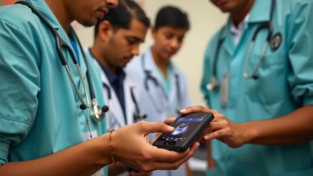
In a 228-bed public hospital in western Guatemala, where diagnostic imaging has long been limited to plain X-rays and heavy workloads crowd out formal teaching, a team of physicians has shown that medical students can learn point-of-care ultrasound (POCUS) not in a classroom, but on the wards, during daily rounds. The program, described in the Journal of General Internal Medicine, embedded handheld ultrasound into the routine clinical work of eight final-year medical students at Hospital Nacional de Coatepeque, and the results offer a technical blueprint for how one of medicine’s most powerful bedside tools might reach the places that need it most.
Point-of-care ultrasound is a compact diagnostic technology: a handheld probe, often paired with a tablet or smartphone, transmits high-frequency sound waves into the body and renders real-time images of fluid collections, organs, and anatomy at the bedside. Unlike a conventional radiograph, which requires a machine, a technician, and a trip to the imaging suite, POCUS can be performed by the clinician examining the patient, in seconds. In hospitals where imaging is limited to plain radiography, the technology effectively collapses the distance between the clinical question and the diagnostic answer. It is also an experiential learning tool, letting trainees see anatomy and pathology directly rather than inferring it from a stethoscope.
Yet despite consensus recommendations from groups in Canada, Europe, and the United States calling for POCUS training in internal medicine, no rounds-integrated curriculum for internal medicine had ever been described in Latin America, and most existing programs fall short of practice-based bedside assessment. The Guatemalan program, led by Dr. Philip K. Angelides of the University of Colorado School of Medicine together with Guatemalan and international collaborators, was designed to close both gaps at once.
The curriculum combined three technical components. First, students completed flipped-classroom prework, studying ultrasound principles and image interpretation before hands-on sessions. Second, they attended a 2-day intensive course focused on pleural and abdominal fluid detection, the core skills for identifying pleural effusions, ascites, and bladder volume. Third, and most importantly, they entered a 2-month longitudinal program in which POCUS was embedded into daily rounds, so that every ward encounter became an opportunity to scan, interpret, and receive feedback in real time.
The program used Butterfly iQ+ handheld ultrasound probes, donated for the educational initiative, paired with Butterfly Cloud, a cloud-based platform where students uploaded their scans for portfolio review by faculty. Feedback flowed through WhatsApp, the messaging platform already ubiquitous in Guatemala, allowing instructors to critique images asynchronously and students to ask questions between rounds. The program also designated three of the eight learners as local champions in a train-the-trainer model, empowering them to teach peers and sustain the program after visiting faculty departed.
Evaluation showed that knowledge and confidence gains largely persisted at 2 months, a finding that matters because competency retention in ultrasound training is notoriously difficult to sustain. Hands-on performance was strongest for pelvic and bladder view acquisition, artifact recognition, and image optimization, skills that involve relatively fixed anatomy and reproducible technique. Performance was lowest for spine sign recognition, the sonographic finding of vertebral bodies appearing in the chest when pleural fluid accumulates behind the lung, and for probe orientation and manipulation, the motor skills that require precise hand-eye coordination and spatial reasoning.
That pattern of strengths and weaknesses is technically revealing. Fluid detection in the bladder and abdomen involves identifying anechoic, or black, collections against relatively predictable landmarks, making it accessible to novices. Spine sign, by contrast, requires understanding the relationship between the lung, the pleural space, and the thoracic spine in three dimensions, and probe manipulation requires the kind of fine motor control that develops only with repeated scanning. The findings align with a growing body of evidence from novice learners showing that cognitive gains in ultrasound come quickly, while motor skill retention follows longer learning curves.
Barriers included probe scarcity and limited institutional support, constraints familiar from POCUS programs across low- and middle-income countries. The champions emerged as key drivers of peer learning, suggesting that the train-the-trainer model may be as important as the technology itself in sustaining programs where faculty time and equipment are limited. The program was reviewed and deemed exempt by the Colorado Multiple Institutional Review Board, and all participants provided informed consent.
The implications reach beyond Guatemala. POCUS has been shown to influence clinical decision-making in emergency departments in Tanzania, to build diagnostic capacity in Rwanda, Malawi, Kenya, and Ghana, and to support training programs in Oman and across Latin America. The Guatemalan study adds a specific technical model: rounds-integrated training, cloud-based portfolio review, messaging-platform feedback, and local champion development, all feasible in a resource-limited internal medicine setting. The authors conclude that rounds-integrated POCUS training is feasible in resource-limited settings and may support early skill development when paired with longitudinal mentorship and local champion development.
For a hospital where imaging has been limited to plain radiography, the arrival of handheld ultrasound represents a shift in what a physical examination can reveal. For medical education, it suggests that the technology need not wait for residency or fellowship, and that the wards themselves, even in hospitals with scarce resources, can become the classroom. The study was published on 8 September 2026 in the Journal of General Internal Medicine.
Subject of Research: POCUS for the People: A Rounds-Integrated Point-of-Care Ultrasound Curriculum for Medical Students at a Guatemalan Public Hospital
Article Title: POCUS for the People: A Rounds-Integrated Point-of-Care Ultrasound Curriculum for Medical Students at a Guatemalan Public Hospital
Article References: Angelides, P. K., León, R., Stelmach, A., Ortiz-López, C., Sánchez, H. A. H., Bolaños, G., Fleshner, M., Sawaya, A., Gamboa, J. E., Hanna, R. M., & Olson, D. (2026). POCUS for the People: A Rounds-Integrated Point-of-Care Ultrasound Curriculum for Medical Students at a Guatemalan Public Hospital. Journal of General Internal Medicine. https://doi.org/10.1007/s11606-026-10797-6
Image Credits: AI Generated
DOI: 10.1007/s11606-026-10797-6
Keywords: point-of-care ultrasound, medical education, global health, Guatemala, POCUS, resource-limited settings, People, Rounds-Integrated, Point-of-Care, Ultrasound, Curriculum, Medical
Cite Scienmag News
Ophelia Keating. (October 17, 2026). Pocket Ultrasound Brings Bedside Imaging to Guatemalan Medical Students. Scienmag. https://scienmag.com/pocket-ultrasound-brings-bedside-imaging-to-guatemalan-medical-students/
Ophelia Keating. "Pocket Ultrasound Brings Bedside Imaging to Guatemalan Medical Students." Scienmag, 17 October 2026, https://scienmag.com/pocket-ultrasound-brings-bedside-imaging-to-guatemalan-medical-students/. Accessed 17 October 2026.
Ophelia Keating. "Pocket Ultrasound Brings Bedside Imaging to Guatemalan Medical Students." Scienmag. October 17, 2026. https://scienmag.com/pocket-ultrasound-brings-bedside-imaging-to-guatemalan-medical-students/


