


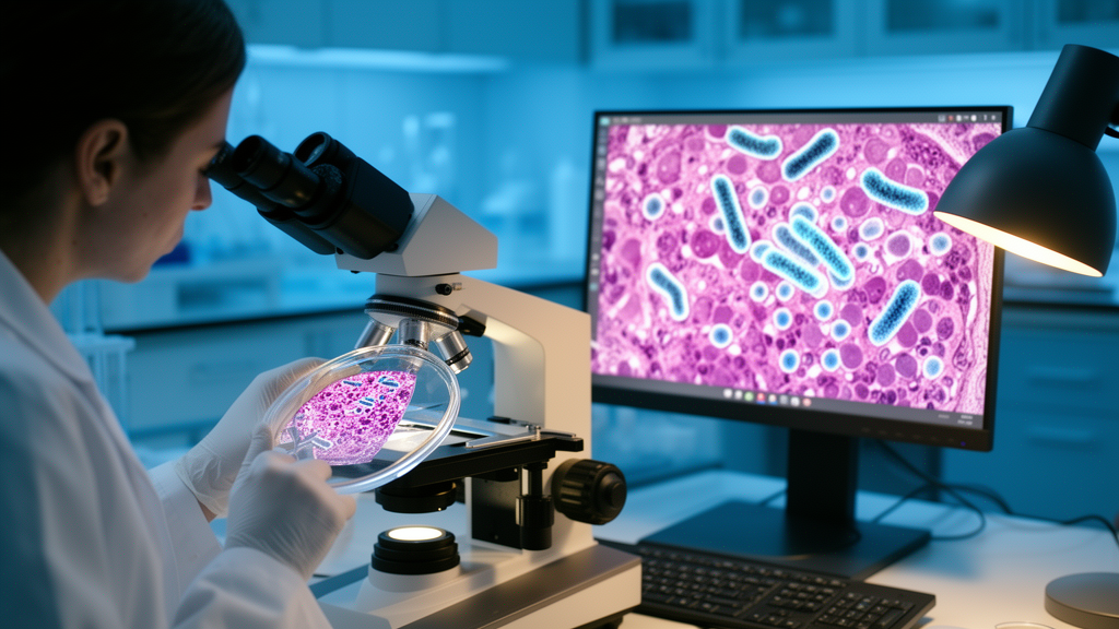
In a finding that blurs the line between microbiology and artificial intelligence, researchers have shown that deep learning models can detect the presence of an intratumoral bacterium directly from routine histopathology slides. The study, published in BMC Medical Imaging, demonstrates that standard hematoxylin and eosin (H&E) stained whole-slide images of colorectal cancer tissue contain latent morphological signatures predictive of Fusobacterium colonization, a microbial genus closely associated with colorectal cancer progression and molecular heterogeneity.
The research team, led by corresponding authors Liping Sun of China Medical University and Huakang Tu of Zhejiang University School of Medicine, set out to determine whether digital pathology slides could serve as a window into the tumor microbiome. Current sequencing-based approaches for detecting intratumoral microbes lack spatial histopathological context, meaning they can confirm a microbe’s presence but cannot reveal where in the tissue it resides or what morphological features accompany its colonization. By contrast, the deep learning approach developed here preserves that spatial dimension while requiring only the slides that pathologists already produce every day.
To test their hypothesis, the investigators assembled an unusually large and diverse patient population: 1,301 colorectal cancer patients drawn from four independent cohorts. These included 608 patients from the TCGA-CRC dataset, 102 from CPTAC-COAD, 298 from the Ningbo Clinical Pathology Diagnosis Center (NCPDC), and 293 from China Medical University (CMU). The primary microbial labels were derived from metagenomic sequencing, with additional label-sensitivity analyses performed using relative abundance thresholds of 0.001% and 0.01% to ensure the findings were not artifacts of a single labeling convention.
On the computational side, the team evaluated multiple pathology foundation models, including CONCH, Gigapath, UNI, UNI2, and Virchow_v2, for predicting the Fusobacterium genus and five representative species from H&E whole-slide images. These foundation models, pretrained on vast collections of pathology images, encode subtle visual patterns that human observers may not consciously register. Among single models, UNI and Virchow_v2 demonstrated the most robust performance, with the highest area under the curve (AUC) reaching 83.0% for Fusobacterium massiliense, one of the five species examined.
The researchers did not stop at single models. They constructed ensemble models using an averaging fusion strategy, combining the outputs of multiple foundation models to improve reliability. The ensembles showed consistently robust performance and high inter-model agreement across different Fusobacterium species, particularly for Fusobacterium nucleatum, where agreement reached 99.2%. Model performance and ensemble robustness were evaluated using classification metrics, agreement analysis, and Shapley value attribution, a technique borrowed from game theory that quantifies each model’s contribution to the final prediction.
Beyond raw predictive accuracy, the study revealed biologically meaningful patterns. Predicted Fusobacterium colonization was consistently enriched in mucinous adenocarcinoma and microsatellite instability (MSI) tumors across multi-cohort association analyses, suggesting that the models were capturing genuine histomorphological niches rather than spurious correlations. These associations align with known clinicopathological features of Fusobacterium-positive colorectal cancers reported in prior sequencing-based studies, lending external validity to the deep learning predictions.
Perhaps the most striking technical contribution came from Grad-CAM analysis, a visualization method that highlights which image regions drive a model’s predictions. The analysis revealed that microbial-associated signals were mainly concentrated in tumor gland lumens and tumor-stroma interface regions, suggesting distinct histomorphological contexts associated with microbial colonization. Multiple-instance learning attention heatmaps and representative pathological review further indicated that high-attention regions frequently involved tumor-stroma interfaces, inflammatory cell-rich areas, mucinous components, and luminal debris or necrosis-like material, painting a recurrent morphological picture of where Fusobacterium-related labels reside in the tissue.
The implications extend well beyond colorectal cancer. The findings demonstrate that routine histopathology contains latent spatial signals associated with intratumoral microbial ecology, highlighting the potential of foundation model-based computational pathology for microbiome characterization and biomarker discovery. Because H&E slides are produced universally and archived in hospitals worldwide, the approach could in principle be applied retrospectively to millions of existing specimens, enabling large-scale studies of tumor-microbe interactions without new tissue collection or sequencing costs.
For clinical practice, the ability to predict intratumoral Fusobacterium from digital slides could support risk stratification and treatment planning. Fusobacterium-positive colorectal cancers have been linked in prior research to distinct molecular features and potentially different responses to therapy, and a slide-based proxy for microbial status could complement existing biomarkers. The authors note that the models were trained and validated across four independent cohorts, providing a degree of generalizability that single-center studies often lack.
The study complies with the Declaration of Helsinki, with ethics approval from Zhejiang University School of Public Health, the Ningbo Clinical Pathology Diagnosis Center, and the First Affiliated Hospital of China Medical University. Written informed consent was obtained from all participants, and analyses involving TCGA and CPTAC datasets used publicly available, de-identified data. The work was supported by the National Natural Science Foundation of China, the National Key R&D Program of China, and the Healthy Zhejiang One Million People Cohort. As foundation models continue to mature, the ability to read microbial ecology from stained tissue may become a standard tool in the computational pathologist’s arsenal, transforming what was once invisible into actionable insight.
Subject of Research: Deep learning prediction of intratumoral Fusobacterium from digital colorectal cancer H&E slides
Article Title: Deep learning prediction of intratumoral Fusobacterium from digital colorectal cancer H&E slides
Article References: Xiang, H., Hu, L., Xu, J., Cai, H., Lu, K., You, Z., Shang, B., Huang, Q., Su, G., Zhang, H., Dai, W., Chang, X., Zhang, Z., Sun, L., & Tu, H. (2026). Deep learning prediction of intratumoral Fusobacterium from digital colorectal cancer H&E slides. BMC Medical Imaging. https://doi.org/10.1186/s12880-026-02696-3
Image Credits: AI Generated
DOI: 10.1186/s12880-026-02696-3
Keywords: Deep, learning, prediction, intratumoral, Fusobacterium, digital, colorectal, cancer, slides, scientific research
Cite Scienmag News
Nathaniel Bowman. (October 17, 2026). AI Reads Bacteria Hidden in Colorectal Cancer Slides. Scienmag. https://scienmag.com/ai-reads-bacteria-hidden-in-colorectal-cancer-slides/
Nathaniel Bowman. "AI Reads Bacteria Hidden in Colorectal Cancer Slides." Scienmag, 17 October 2026, https://scienmag.com/ai-reads-bacteria-hidden-in-colorectal-cancer-slides/. Accessed 17 October 2026.
Nathaniel Bowman. "AI Reads Bacteria Hidden in Colorectal Cancer Slides." Scienmag. October 17, 2026. https://scienmag.com/ai-reads-bacteria-hidden-in-colorectal-cancer-slides/


