


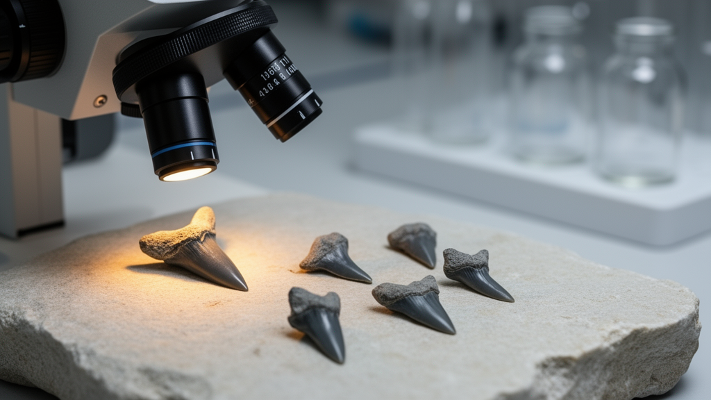
Deep in the limestones of south-western China, the teeth of long-extinct sharks are rewriting what scientists know about the dawn of modern shark lineages. A new taxonomic study published in the Swiss Journal of Palaeontology describes nine fossil shark teeth from the Ladinian–Carnian interval of the Zhuganpo Member of the ‘Falang’ Formation in Guizhou and Yunnan Provinces, South China. The teeth belong to four euselachian sharks: one of uncertain affinity, two indeterminate neoselachians and one potential hexanchid. Although the specimens are tiny, some measuring barely a few tenths of a millimetre in height, they carry enormous systematic weight, because they document, for the first time, the three-dimensional dental vascularisation of Triassic chondrichthyans.
The teeth were recovered from two sections that have become famous among vertebrate palaeontologists. The Nimaigu section in Wusha Town, Xingyi City, south-western Guizhou Province, and the Yize section in Changdi Town, Luoping County, eastern Yunnan Province, both lie on the south-western part of the Yangtze Carbonate Platform. During the Triassic, these sites recorded an upward transition from shallow-water dolomite of the Yangliujing Formation to deep-water marl and nodular limestone of the Zhuganpo Member, and subsequently to the black shale and marl of the Wayao Member, which represents the maximum flooding surface. The ‘Falang’ Formation is renowned for yielding two exceptionally well-preserved marine vertebrate faunas: the latest Ladinian Xingyi Fauna and the early Carnian Guanling Biota, both famous for their marine reptiles and fishes.
Fieldwork in 2017, 2018 and 2019 produced large quantities of rock samples from the ‘Falang’ Formation at both sections. The samples were broken into fragments approximately four centimetres in size and dissolved using ten percent acetic acid, a standard technique for freeing microfossils from carbonate rock. Conodonts and chondrichthyan teeth were then manually picked from the residues under a stereomicroscope. Nine shark teeth were examined in total: specimen GMPKU-P-4268 from the Yize section and GMPKU-P-4261 to 4267 and 4269 from the Nimaigu section. All specimens are housed in the Geological Museum of Peking University in Beijing.
The teeth were photographed with a JEOL JCM-6000 Plus scanning electron microscope under an acceleration voltage of 10 kV at the School of Earth and Space Sciences, Peking University. Two specimens, GMPKU-P-4263, assigned to Euselachii gen. et sp. indet., and GMPKU-P-4266, assigned to Neoselachii gen. et sp. indet. 1, were scanned with a SkyScan 1172 micro-CT device at the State Key Laboratory of Systematic and Evolutionary Botany, Institute of Botany, the Chinese Academy of Sciences. The scans used a 180-degree rotation, a rotation step of 0.2 degrees and a pixel size of 0.56 micrometres, with the source voltage and current set to 48 kV and 200 microamperes. The teeth and their vascular systems were then reconstructed in three dimensions using AVIZO 2022.2, revealing internal structures that no external examination could ever expose.
Euselachii gen. et sp. indet. is represented by five isolated teeth from the late Ladinian layers of the Nimaigu section. The teeth are elongated mesiodistally and narrow labiolingually, measuring 0.21 to 0.37 millimetres in height and 0.58 to 1.10 millimetres mesio-distally. A gradient monognathic heterodonty is evident: anterolateral teeth have an arched root and relatively separated cusps, whereas posterolateral teeth display a straight root and fused cusps. The crown carries a robust pyramidal main cusp flanked by two to four lateral cusplets on each side, with one or two significantly smaller intermediate cusplets between the main cusp and the first pair of lateral cusplets. All cusps are inclined distally and curved lingually, and adjacent cusps are separated by V-shaped notches on anterolateral teeth but fused at their base on posterolateral teeth.
Micro-CT scanning of specimen GMPKU-P-4263 revealed an orthodont histotype. The crown consists of enameloid and a compact layer of underlying orthodentine, which enfolds a large longitudinal vascular canal at the crown-root junction, but no ascending pulp cavity can be seen. Osteodentine is restricted to the root. The vascular system comprises a cylindrical longitudinal vascular canal with a diameter of 30 to 90 micrometres extending along the crown-root junction, a single-layered vascular plexus of interconnected canals ranging from 8 to 40 micrometres in diameter forming numerous circular grids, short connective vascular canals linking the plexus to the overlying longitudinal canal, and ascending vascular canals. At the distal extremity, the longitudinal canal branches into three thinner canals. This longitudinal canal, unaccompanied by an ascending pulp cavity, also occurs in other non-neoselachian euselachians with very different crown shapes, from the flat-crowned hybodont Palaeobates angustissimus to the multicuspid Lamarodus triangulus, suggesting that its presence may be independent of tooth morphology.
Neoselachii gen. et sp. indet. 1 is represented by a single broken tooth, GMPKU-P-4266, from layers 145 to 146 of the Nimaigu section, dated to the late Ladinian. The tooth measures 0.75 millimetres in height and 1.43 millimetres mesio-distally, with a robust biconvex main cusp curved lingually and slightly inclined distally, flanked by one lateral cusplet on the mesial side and two on the distal side. The labial face of the main cusp is devoid of ornamentation, while the lingual face bears seven faint vertical ridges. The root is low, kidney-shaped in basal view, and protrudes lingually. Its vascularisation may represent a transitional state between anaulacorhize and polyhemiaulacorhize types.
The internal architecture of this neoselachian proved even more remarkable. Micro-CT reconstruction showed an orthodont tooth in which a reduced pulp cavity ascends into the main cusp, while porous osteodentine is confined to the root. The vascular system contains two longitudinal vascular canals, each 130 to 150 micrometres in diameter and nearly as long as the tooth itself, interconnected by short vertical or oblique connective canals. Four parallel main transverse vascular canals, 52 to 93 micrometres in diameter, run labiolingually through the middle of the root, accompanied by shorter and narrower secondary transverse canals. The vascularisation resembles that of Synechodus sp. from the Early Cretaceous of northern France, which also shows a longitudinal vascular canal, ascending pulp cavity and transverse canals, but the Chinese specimen differs by possessing two large longitudinal canals rather than one significantly narrower canal, and by lacking the meshed secondary vascular canals of Synechodus.
The remaining two taxa are known from single or paired teeth. Neoselachii gen. et sp. indet. 2 is represented by one anterior tooth, GMPKU-P-4267, from layer 186 of the Nimaigu section, bearing a slender main cusp without lateral cusplets, two sharp lateral cutting edges and a low bilobate root with a lingually unroofed median canal, a vascularisation reminiscent of the hemiaulacorhize type. The specimen recalls an undetermined neoselachian from the Ladinian–Carnian interval of Guanling, Guizhou, and the two could belong to the same species, although the Guanling tooth is significantly larger. Hexanchidae? gen. et sp. indet. is represented by two broken teeth, GMPKU-P-4268 and 4269, with labiolingually compressed crowns bearing a triangular main cusp flanked by three pairs of smaller lateral cusplets that decrease in height laterally. Because the root is missing, the affiliation remains tentative, but the crown morphology points to the cow shark family Hexanchidae, comparable with material from the late Permian of Japan and the middle Jurassic of England.
Stratigraphically, Hexanchidae? gen. et sp. indet. is the only taxon that may range across the Ladinian–Carnian boundary, with one tooth from late Ladinian layers at Nimaigu and another from early Carnian strata at Yize, above the first occurrences of the conodonts Quadralella polygnathiformis and Quadralella intermedia. The remaining three taxa are confined to the late Ladinian at Nimaigu. Palaeoecologically, the four taxa display non-durophagous feeding behaviour: Euselachii gen. et sp. indet. exhibits a grasping-swallowing dentition with cladodont-like crowns, Neoselachii gen. et sp. indet. 1 may exhibit a grasping type tooth, Neoselachii gen. et sp. indet. 2 displays a tearing type tooth with a slender main cusp and sharp cutting edges, and Hexanchidae? gen. et sp. indet. bears cutting type teeth. These findings confirm that the Ladinian–Carnian shark assemblage at Nimaigu lacks durophagous forms, making it different from other contemporaneous shark faunas worldwide that were predominantly represented by durophagous sharks. With the new material, at least nine chondrichthyan taxa have now been found at the Nimaigu section, and all four newly described chondrichthyans appear to be endemic, unknown outside South China, underscoring how much of the early evolution of euselachian sharks remains hidden in the world’s Triassic rocks.
,
Subject of Research: A taxonomic and micro-CT study of nine euselachian shark teeth from the Ladinian–Carnian Zhuganpo Member in Guizhou and Yunnan Provinces, South China, describing four taxa and revealing Triassic dental vascularisation in 3D.
Article Title: New euselachian teeth from the Ladinian–Carnian interval of Guizhou and Yunnan Provinces, South China
Article References: Zhao, S., Li, J., Cuny, G., & Sun, Z. (2026). New euselachian teeth from the Ladinian–Carnian interval of Guizhou and Yunnan Provinces, South China. Swiss Journal of Palaeontology, 145, 313-326. https://doi.org/10.3897/sjp.145.185785
Image Credits: AI Generated
Keywords: Triassic, chondrichthyans, palaeontology, micro-CT, South China, shark teeth, euseleachii, neoselachians, hexanchids, dental histology, euselachian, teeth
Cite Scienmag News
Gavin Prescott. (October 17, 2026). Ancient shark teeth from South China reveal hidden Triassic diversity in 3D. Scienmag. https://scienmag.com/ancient-shark-teeth-from-south-china-reveal-hidden-triassic-diversity-in-3d/
Gavin Prescott. "Ancient shark teeth from South China reveal hidden Triassic diversity in 3D." Scienmag, 17 October 2026, https://scienmag.com/ancient-shark-teeth-from-south-china-reveal-hidden-triassic-diversity-in-3d/. Accessed 17 October 2026.
Gavin Prescott. "Ancient shark teeth from South China reveal hidden Triassic diversity in 3D." Scienmag. October 17, 2026. https://scienmag.com/ancient-shark-teeth-from-south-china-reveal-hidden-triassic-diversity-in-3d/


