


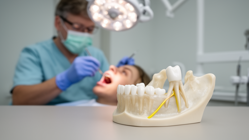
Oral surgeons have long faced a dilemma when a lower wisdom tooth sits dangerously close to the inferior alveolar nerve, the sensory highway that carries feeling from the lower lip, chin, and teeth to the brain. Extracting such a tooth risks permanent numbness; leaving it risks recurrent infection. A newly documented case from Uppsala, Sweden, offers a striking illustration of how a nerve-sparing technique called coronectomy can work — and how the body itself may help the surgeon by moving retained tooth roots away from the nerve over time.
The case, published in the journal Clinical Case Reports, involved a 41-year-old woman referred for recurrent pericoronitis, an inflammatory condition around a partially erupted lower left third molar. Imaging revealed an anatomical rarity: the tooth had four roots, and the inferior alveolar nerve passed directly between them, visibly compressed by the root structure. According to prior research cited in the report, this configuration occurs in roughly 1 percent of cases with open roots and about 0.4 percent with fused apices.
Faced with this anatomy, surgeons at the Department of Oral and Maxillofacial Surgery chose coronectomy — the deliberate removal of only the crown while leaving the roots in place. The procedure, performed in May 2019 under local anesthesia with mild sedation, aimed to eliminate the source of recurrent infection while avoiding direct trauma to the nerve. The surgeons noted a technical shortcoming: a small horn of enamel remained adjacent to the second molar, a detail that would prove relevant years later.
What happened next illustrates a biological phenomenon well known but rarely seen so dramatically. Over the following years, cone-beam CT scans showed that the retained root complex had migrated superiorly, distally, and lingually — moving away from the nerve, which now lay inferior to the roots. Root migration after coronectomy occurs in up to 50 percent of cases, typically within the first two years, and is generally considered a favorable response as bone remodels around the retained structure.
Five years after the initial procedure, however, the patient returned with new symptoms. The migrated roots had become associated with recurrent infection, and imaging showed bone loss around the root apices. Surgeons performed a delayed root removal in April 2024, retrieving the fused lingual root complex as a single specimen and the buccal root in two fragments. Healing proceeded without complication.
The case highlights both the promise and the limits of coronectomy. By preserving the roots initially, surgeons avoided nerve injury in a patient whose anatomy made conventional extraction extraordinarily risky. But the technique is not without long-term consequences: retained roots can migrate, become exposed, or develop secondary infection years after the initial procedure. The report’s authors emphasize that long-term clinical and radiological follow-up is essential, particularly when technical imperfections such as retained enamel are present.
Coronectomy has gained acceptance as a valuable alternative for high-risk lower third molars. Studies show significantly shorter duration of postoperative pain compared with full extraction, and lower rates of temporary nerve disturbance. But the procedure requires careful patient selection: active infection, extensive caries, or immunocompromise may contraindicate it. The Uppsala team notes that evidence-based follow-up protocols remain poorly defined, and more research is needed to identify factors influencing root migration and long-term outcomes.
For now, the case stands as a vivid example of surgical pragmatism — and of the body’s capacity to assist in its own repair. When a wisdom tooth’s roots threaten a nerve, sometimes the wisest course is to let biology do the moving.
The biological mechanism behind root migration after coronectomy remains incompletely understood, but several explanations have been proposed in the literature. One leading hypothesis holds that the retained roots, once relieved of occlusal load from the removed crown, are gradually pushed upward by the remodeling forces of the surrounding alveolar bone, much like an ankylosed structure being slowly elevated during growth-related bone turnover. Another view emphasizes the role of the periodontal ligament remnants and the follicular tissues that remain attached to the retained root complex, which may retain some of the eruptive potential that originally drove tooth eruption during adolescence. Whatever the precise mechanism, the practical consequence is consistent across published series: the roots tend to drift toward and sometimes above the level of the alveolar crest, and in doing so they frequently move away from the inferior alveolar canal, increasing the bony separation between nerve and dentin.
The timing of this migration matters clinically. The source report notes that migration is most active during the first six months after surgery, with mean displacement figures in the range of roughly two to five millimeters within the first year, and that the process generally plateaus by the second or third year of follow-up. In the Uppsala case, the movement continued to be radiographically evident years later, with the root complex ultimately coming to lie above the mandibular canal rather than straddling it. This repositioning is precisely what makes delayed removal of the roots far safer than primary extraction would have been: by the time surgical removal became necessary in 2025, the nerve no longer interposed itself between the roots, and the operation could proceed without the extreme nerve-injury risk that had characterized the original anatomy.
Age and sex appear to modulate the phenomenon. The cited literature suggests that migration is more pronounced in younger patients, plausibly because bone turnover and remodeling capacity decline with age, and that it tends to be somewhat greater in females. At forty-one, the patient in this case sat in a middle range, yet her migration was substantial enough to alter the surgical calculus entirely. Clinicians counseling patients about coronectomy can therefore reasonably describe migration as the expected outcome rather than an incidental curiosity, occurring in the large majority of cases according to the figures cited in the report.
The delayed complication in this case also deserves attention from a diagnostic standpoint. Five years after the initial surgery, the patient presented with swelling, purulent discharge, and a fistula located distobuccal to the second molar — a presentation that could easily be mistaken for a recurrent pericoronal problem or an endodontic lesion of the adjacent tooth. Cone-beam CT proved decisive, revealing periodontal bone loss on the mesial aspect of the retained root complex and apical periodontitis, confirming that the retained roots themselves had become the infectious focus. This underscores a practical point for clinicians: when a patient with a prior coronectomy returns with unexplained symptoms in the retromolar region years after surgery, advanced imaging should be considered early, since two-dimensional panoramic radiography may not adequately demonstrate periapical pathology or the precise relationship between migrated roots and the canal.
It is also worth noting the surgical implications of the migration itself. When the root complex was finally removed, the operating team observed bone formation in the interradicular area — new bone had grown into the space between the original roots even in the presence of infection. This bony infilling is a hallmark of successful coronectomy outcomes and reflects the same remodeling process that drives migration. The fused lingual root complex was retrieved as a single specimen, while the buccal root fractured during elevation and was removed in two fragments, together accounting for all four original anatomical roots. The specimen photographs published with the case provide a rare direct visual record of the anatomy that preoperative imaging had only suggested.
The question of how long retained roots should be monitored has no settled answer, and this case adds to the argument that follow-up should extend well beyond the conventional one- or two-year window. Most published series focus on migration distances and nerve outcomes during the first two years, after which many patients are discharged. Yet the complication here emerged at the five-year mark, long after routine surveillance would typically have ended. Whether the retained enamel horn noted on the immediate postoperative radiograph contributed to the late infection is impossible to establish with certainty from a single case, but residual enamel is listed among the recognized contraindications and technical pitfalls of the procedure precisely because it may permit bacterial ingress or cyst formation. Surgeons performing coronectomy should therefore verify complete enamel removal intraoperatively and document the result radiographically before closure.
Finally, the case illustrates the value of cone-beam computed tomography at both ends of the treatment timeline. Preoperatively, the three-dimensional imaging revealed the four-root anatomy and the interposition of the mandibular canal between the roots — findings that panoramic radiography alone could not have resolved with sufficient confidence to plan a coronectomy. Postoperatively, the same modality documented the migration vector, the new position of the nerve relative to the root apices, and the extent of periapical bone loss that justified delayed removal. As access to CBCT expands in oral surgical practice, its role in both selecting candidates for coronectomy and managing their long-term follow-up is likely to grow, particularly for anatomically complex cases such as this one, where the margin between a routine extraction and a permanent sensory deficit was measured in fractions of a millimeter.
Subject of Research: Coronectomy of a mandibular third molar with the inferior alveolar nerve passing between the roots, documenting root migration away from the nerve and delayed root removal after secondary infection.
Article Title: Root Migration After Coronectomy of a Mandibular Third Molar With the Inferior Alveolar Nerve Passing Between the Roots: A Case Report
Article References: Marquez Rocco, V., Thor, A., Hashim, A. A., & Bellerud, D. (2026). Root Migration After Coronectomy of a Mandibular Third Molar With the Inferior Alveolar Nerve Passing Between the Roots: A Case Report. Clinical Case Reports, 14(10), Article e73673. https://doi.org/10.1002/ccr3.73673
Image Credits: AI Generated
DOI: 10.1002/ccr3.73673
Keywords: coronectomy, inferior alveolar nerve, mandibular third molar, root migration, oral surgery, pericoronitis, nerve injury prevention, Root, Migration, Mandibular, Third, Molar
Cite Scienmag News
Ophelia Keating. (October 17, 2026). When Wisdom Tooth Roots Move Away From the Nerve: A Rare Coronectomy Case. Scienmag. https://scienmag.com/when-wisdom-tooth-roots-move-away-from-the-nerve-a-rare-coronectomy-case/
Ophelia Keating. "When Wisdom Tooth Roots Move Away From the Nerve: A Rare Coronectomy Case." Scienmag, 17 October 2026, https://scienmag.com/when-wisdom-tooth-roots-move-away-from-the-nerve-a-rare-coronectomy-case/. Accessed 17 October 2026.
Ophelia Keating. "When Wisdom Tooth Roots Move Away From the Nerve: A Rare Coronectomy Case." Scienmag. October 17, 2026. https://scienmag.com/when-wisdom-tooth-roots-move-away-from-the-nerve-a-rare-coronectomy-case/


