


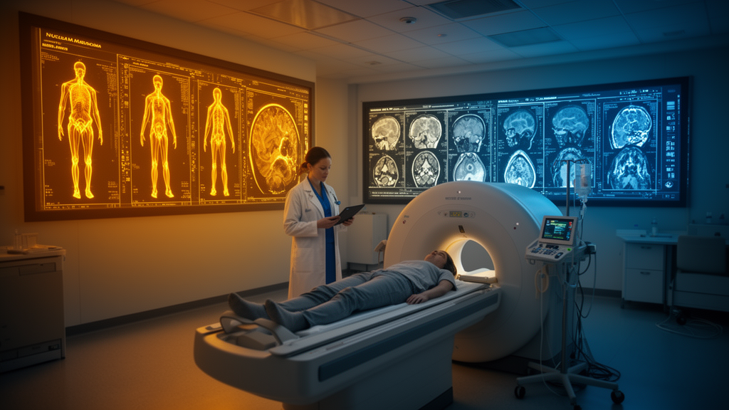
Imagine walking into a nuclear medicine clinic, receiving a single radioactive injection, and walking out with not one but two molecular portraits of your cancer. That tantalizing possibility has moved a step closer to reality, thanks to a team of researchers in Xi’an, China, who have trained an artificial intelligence system to generate one type of PET scan from another. In a study published in Frontiers in Nuclear Medicine, the group describes a cascaded deep learning framework that synthesizes 18F-FDG PET images — the workhorse of oncological imaging — from 18F-octreotide PET scans, which are used to detect neuroendocrine tumors. The results, while preliminary, hint at a future where patients could receive a comprehensive dual-parameter molecular workup with half the injections, half the scans, and half the radiation dose.
The clinical motivation is rooted in a well-known biological dilemma. Neuroendocrine tumors, which most commonly arise in the gastroenteropancreatic system and lungs, are notoriously heterogeneous. Somatostatin receptor imaging with 18F-octreotide, which binds to overexpressed SSTR2 receptors on tumor cells, is the cornerstone of diagnosis. But receptor expression alone tells an incomplete story. Roughly 15 to 30 percent of neuroendocrine tumor patients show so-called SSTR–FDG discordance: tumors that light up on octreotide scans but not on FDG scans tend to be well-differentiated and indolent, and are prime candidates for peptide receptor radionuclide therapy. The reverse pattern — FDG-positive but receptor-negative — signals dedifferentiation, aggressive biology, and poor prognosis, calling instead for chemotherapy or targeted agents. Dual-positive lesions demand integrated interpretation. In short, clinicians need both tracers to stage the disease accurately and choose the right treatment.
The problem is that getting both tracers traditionally means two separate PET/CT examinations on different days. Each whole-body FDG PET/CT carries an effective dose of roughly 14 millisieverts, so doubling the scans doubles the radiation burden. Patients must return days later to avoid tracer interference, costs mount, and some patients simply drop out of the workflow altogether. An AI system that could conjure a convincing FDG image from an octreotide scan would collapse this two-step process into one — a single-injection, dual-parameter imaging paradigm that the authors argue could be especially valuable for patients requiring lifelong surveillance, such as those with multiple endocrine neoplasia type 1.
What makes this synthesis genuinely difficult is that the two tracers probe fundamentally different biology. FDG tracks glucose metabolism; octreotide tracks receptor expression. Their lesion contrast mechanisms, physiological background distributions, and standardized uptake value (SUV) dynamic ranges differ intrinsically. Prior work has shown that single-stage generative networks struggle with such cross-domain translation: they tend to couple the coarse mapping between image domains with fine-detail reconstruction in a single feature space, and in minimizing overall loss they adopt conservative strategies that blur or over-smooth metabolically hot regions. Studies of MRI-to-PET synthesis have found that errors balloon precisely in regions of high tracer uptake — exactly the regions that matter most for diagnosis.
The Xi’an team’s answer is a cascaded conditional generative adversarial network built on the Pix2Pix framework, with two sequential generator–discriminator pairs. The first stage, a 3D U-Net, performs anatomically constrained coarse synthesis: it establishes organ localization and plausible background uptake, solving what the authors call the presence problem. The second stage, a self-attention 3D V-Net with residual learning, refines FDG-specific metabolic features, correcting SUV bias and restoring lesion contrast — the accuracy problem. A self-attention module at the deepest encoder level reshapes feature maps into sequential representations and computes attention weights through Query, Key, and Value embeddings, allowing the network to capture long-range spatial dependencies that improve anatomical consistency and lesion boundary delineation. Both discriminators use multi-scale 3D PatchGAN architectures that judge authenticity at three spatial resolutions simultaneously.
The researchers trained and tuned this machinery on paired scans from 71 tumor patients who underwent both FDG and octreotide PET/CT between May 2022 and November 2025, splitting the data at the patient level into training, validation, and test cohorts in a 6:2:2 ratio. Two engineering choices proved decisive. First, preprocessing: because the tracers’ SUV distributions differ so markedly, the team compared logarithmic transformation, Z-score normalization, and a combined approach. The combination won decisively, achieving a structural similarity index (SSIM) of 0.8454 in the 14-patient held-out test set, versus 0.7831 for normalization alone and 0.7438 for log transformation alone, with all five objective metrics — SSIM, PSNR, MSE, MAE, and NMI — differing significantly across strategies. Logarithmic compression tames the extreme dynamic range that would otherwise let hot lesions dominate gradient updates, while normalization stabilizes convergence.
Second, input parameterization mattered enormously. Comparing two configurations on the identical test cohort, the team found that a larger resampling size of 256 by 256 by 512 voxels (Group 2) dramatically outperformed the baseline 128 by 128 by 256 configuration (Group 1). SSIM rose from 0.7431 to 0.8854, a relative improvement of 19.1 percent, while peak signal-to-noise ratio jumped from 15.34 to 28.15 decibels — an 83.5 percent relative gain. Mean squared error fell by 80.8 percent and mean absolute error by 63.3 percent, all with large effect sizes. The authors are careful to note that the two configurations also differed in training iterations and batch size, so the improvement reflects the overall parameter configuration rather than resampling resolution in isolation; a controlled one-variable-at-a-time ablation is planned as future work.
The most clinically resonant findings concern quantitative fidelity rather than pixel-level similarity. Three blinded nuclear medicine physicians rated the synthetic images a mean of 4.53 on a five-point Likert scale, with all 14 test images scoring above four and ten rated as excellent, and good inter-reader agreement. More importantly, lesion-level analysis showed almost perfect correlations between synthetic and real FDG images: Pearson coefficients of 1.0000 for SUVmax, 0.9995 for SUVmean, and 0.9991 for SUVpeak, with no significant differences on paired t-tests and Bland–Altman biases of no more than 0.073 SUV units, all differences falling within the 95 percent limits of agreement. Liver background uptake, a standard reference organ, was equally consistent, with mean SUVs of 3.39 versus 3.40 and no significant difference.
The limitations, which the authors enumerate candidly, are substantial. The study is a retrospective, single-center pilot with only 71 patients and a 14-patient test set, so the precision of the agreement estimates is limited and generalizability across scanners, reconstruction protocols, and patient populations remains unproven. Only the largest lesion per patient was analyzed, which may overestimate performance since small lesions — five of the test lesions were under 20 millimeters — are the hardest to synthesize owing to partial-volume effects. Synthesis may also falter in dedifferentiated tumors with low receptor expression but high FDG uptake, precisely the biological scenario where the tracers diverge most. The framework uses PET data only, ignoring the anatomical information in the accompanying CT, and the exploratory quadrant analysis of SSTR/FDG concordance categories lacked enough discordant lesions for meaningful subgroup statistics. Crucially, the study demonstrates image synthesis, not diagnostic utility: no diagnostic accuracy, staging, or treatment-decision analyses were performed, and the Likert ratings reflect visual quality rather than diagnostic equivalence.
Even with those caveats, the study stands as one of the first demonstrations of cross-biological tracer translation in whole-body oncological PET, and it offers a practical recipe — cascaded task decoupling, combined log-plus-normalization preprocessing, and higher input resolution — for others attempting the feat. The authors call for prospective, multicenter validation, benchmarking against contemporary generative architectures, small-lesion enhancement strategies, whole-organ SUV calibration, and ultimately studies testing whether synthesized images change staging, therapy selection, or patient outcomes. If those trials succeed, the vision of a single injection yielding a dual-parameter molecular fingerprint of a tumor could transform how neuroendocrine cancer is imaged — cutting radiation, cost, and clinic visits while preserving the complementary biological information that clinicians increasingly rely on.
Subject of Research: Deep learning synthesis of 18F-FDG PET images from 18F-octreotide PET for neuroendocrine tumor imaging
Article Title: Cascaded deep learning for synthesis of 18F-FDG PET from 18F-octreotide: model parameter optimization and performance evaluation
Article References: Chang, R., Yang, L., Gao, J., Liu, X., Wang, B., Li, Y., & Duan, X. (2026). Cascaded deep learning for synthesis of 18F-FDG PET from 18F-octreotide: model parameter optimization and performance evaluation. Frontiers in Nuclear Medicine, 6, Article 1920721. https://doi.org/10.3389/fnume.2026.1920721
Image Credits: AI Generated
DOI: 10.3389/fnume.2026.1920721
Keywords: PET imaging, deep learning, generative adversarial networks, 18F-FDG, 18F-octreotide, neuroendocrine tumors, image synthesis, cross-tracer translation, SUV quantification, nuclear medicine, conditional GAN, radiation dose reduction
Cite Scienmag News
Ophelia Keating. (October 17, 2026). AI Conjures FDG PET Scans From a Single Octreotide Injection, New Study Shows. Scienmag. https://scienmag.com/ai-conjures-fdg-pet-scans-from-a-single-octreotide-injection-new-study-shows/
Ophelia Keating. "AI Conjures FDG PET Scans From a Single Octreotide Injection, New Study Shows." Scienmag, 17 October 2026, https://scienmag.com/ai-conjures-fdg-pet-scans-from-a-single-octreotide-injection-new-study-shows/. Accessed 17 October 2026.
Ophelia Keating. "AI Conjures FDG PET Scans From a Single Octreotide Injection, New Study Shows." Scienmag. October 17, 2026. https://scienmag.com/ai-conjures-fdg-pet-scans-from-a-single-octreotide-injection-new-study-shows/


