


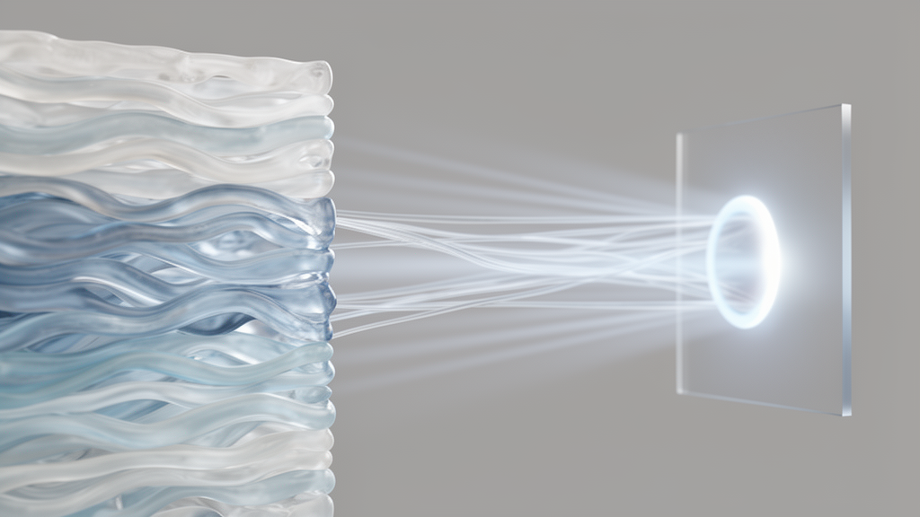
Second harmonic generation (SHG) microscopy has become one of the most powerful label-free tools for studying collagen microstructure in biological tissues. The technique exploits a nonlinear optical process in which two photons at a fundamental frequency interact with a non-centrosymmetric medium and combine to generate a new photon at twice the frequency. Because collagen, microtubules, and myosin filaments exhibit non-centrosymmetric organization similar to a crystal, they generate strong second harmonic signals that can be imaged in detail without staining or fixation. Since the phenomenon was first observed in 1961, when a second harmonic signal at half the wavelength of the incident laser was generated as light passed through a crystal, applications have spanned photonics, optical communications, plasmonics, and biology.
Most SHG research to date has relied on forward-detected signals acquired from ultra-thin tissue sections. Because the forward signal is coherent, the resulting images provide a clear and detailed representation of collagen in the sample under study. However, the requirement for the sensor to sit behind the sample generally limits forward-SHG to ex vivo experiments on thin sections. Backward-detected SHG, by contrast, uses the portion of the signal scattered back from the tissue and therefore holds real potential for translation to in vivo imaging contexts. The problem is that the origin of the backward signal is not fully understood. It represents complex optical interactions, and interpretation of backward images remains challenging, which has led to repeated misinterpretations of the collagen structure of biological tissues.
A team of researchers from University College London and the University of Arizona has now addressed this puzzle in a study published in Frontiers in Photonics. Using a custom optical chamber that allows bidirectional SHG imaging of the cornea under controlled pressure, they assessed the spatial relationship between features in backward images and the underlying structural features of collagen visible in paired forward images. Their central finding is striking: collagen waviness, known as crimp, is a key structural feature responsible for periodic bands of fluctuating signal intensity in backward SHG images. Structures that numerous previous studies had identified as collagen bundles, and even used to compute predominant orientations, are in fact oriented perpendicularly to the actual lamellar direction and originate from signal contributions from different regions of wavy lamellae.
The cornea was selected as the imaging target for several reasons. It is a transparent, collagenous tissue within which the collagen has a specific depth-dependent, highly ordered arrangement that has been characterized previously using histology, scanning electron microscopy, transmission electron microscopy, and X-ray diffraction techniques. The transparent nature of the tissue enables full-depth imaging in both directions of intact tissue, ensuring the structure closely represents the in vivo state, while the complex collagen arrangement and access to previously acquired structural information facilitate convenient validation and a means to assess the origin of imaging artefacts. Ten fresh porcine eyes were obtained from a local abattoir, and corneas were imaged within seventy-two hours postmortem in an intact, non-processed state to prevent structural modifications induced by sectioning or fixation agents.
Imaging was performed with a Zeiss LSM980 on an upright Examiner.Z1 microscope, with laser excitation provided by a Spectra-Physics Insight DS+ laser tuned to 820 nanometers, linearly polarized in the x-axis of the image plane. Initial imaging was performed with the cornea remaining within the eye globe at normal physiological intraocular pressure, using reflection-mode SHG at two magnifications. The cornea was then dissected from the globe with a three-millimeter rim of sclera and re-imaged to acquire simultaneous forward- and backward-detected SHG signals for direct comparison. The custom-designed chamber allowed the cornea to be pressurized with phosphate buffered saline while a manometer provided real-time pressure monitoring, enabling the team to investigate the effects of pressure variation on collagen bundles and the subsequent effect on the generated images. To the best of the authors’ knowledge, this is the first experimental setup described that enables forward imaging of the cornea in its unaltered, pressurized state, providing layer-by-layer detail and the ability to capture structural changes under different loads.
The results revealed a consistent spatial relationship between the two signals. Backward-detected images of the posterior corneal stroma showed highly organized, vertically aligned bands of high-intensity signal with periodic spacing, structures previously interpreted as collagen bundles, while areas of low signal were believed to be regions absent of collagen, corresponding to voids or interlamellar spacing. Higher magnification resolved speckled, elongated patterns oriented perpendicularly, rather than aligned, to the structures previously identified as bundles. When periodic regions of the backward images were overlaid with the corresponding regions of the forward images, the aligned bands in the backward image corresponded to regions of the wavy bundles observed in the forward images. The dark regions did not correspond to voids but to absent or weak backward signal due to the specific collagen position along the wavy lamellae. The forward-detected bundles consistently ran perpendicular to the backward-detected high-signal bands throughout the entire imaging plane.
To demonstrate this mechanism dynamically, the cornea was mounted in the transparent chamber and imaged throughout its depth, first at low pressure of approximately five millimeters of mercury and then at supraphysiological levels of approximately sixty millimeters of mercury, pressures relevant to disease states such as glaucoma and hypotony. It had been theorized, but not yet proven, that collagen waviness is a structural mechanism governing the corneal response to increasing intraocular pressure. At low pressure, the collagen bundles appeared wavy and crimped, with periodic features clearly evident in the backward images. When the pressure was raised, the forward image revealed straightened bundles, while the backward image showed a more uniform forward-to-backward signal and loss of the periodic bands previously observed. In some imaging planes where the collagen did not uncrimp completely, periodic patterns remained in the backward images in regions directly corresponding to the crimped areas in the forward images. A complete layer-by-layer recording across a thickness of 985 micrometers showed the collagen becoming noticeably uncrimped after the pressure increase, with clear differences in the backward signal between crimped and uncrimped states.
The findings also clarify the theoretical frameworks proposed to explain SHG directionality. Theoretical models based on fibril size suggest that fibrils with diameters on the order of the SHG wavelength generate predominantly forward-directed signals, whereas smaller fibrils produce nearly equal forward and backward signals. However, this theory cannot account for the difficulty in correlating backward and forward signals in the cornea, which has a consistent fibril diameter of approximately thirty-one nanometers in pigs across the depth. A more comprehensive quasi-phase matching model treats the fibril no longer as an isolated SHG generator but considers the collective packing of fibrils within a domain, which influences phase-matching conditions and the continuity of coherent second harmonic generation. The team extended this framework by introducing a tilt parameter, defined as the out-of-plane angle between the collagen bundles and the axial propagation direction. This inclination, inherent to the crimp pattern, induces a change in the effective domain size, affecting phase-matching conditions and thus the forward-to-backward signal distribution, creating a periodic alternation between different forward-to-backward ratios that matches the sides of the crimped patterns in the forward images.
The implications extend well beyond correcting image interpretation. A previous study using backward SHG imaging to evaluate corneal response to intraocular pressure changes had observed a narrowing of dark low-signal bands and attributed it to changes in interlamellar spacing. The new study clarifies that changes to these periodic intensity levels occur due to straightening of the crimped lamellae under increased pressures, meaning a key biomechanical mechanism governing the corneal response to pressure was previously overlooked. Because these structural changes can occur within very short timescales, during each heartbeat when the cornea experiences pulsatile intraocular pressure variations, this dynamic behavior must be considered when analyzing backward signals, particularly if an in vivo diagnostic device or AI-based model is to be developed to distinguish between normal and diseased states. The authors note that a key limitation of the work is the absence of quantitative analysis of the correlation between the two signals, since many confounding variables at both the fibrillar and bundle level are expected to affect both signals, but this remains a future aim of the continued work.
With respect to clinical translation, the potential is vast. Diseases such as keratoconus and ectasia are associated with structural and biomechanical changes but are currently not fully understood and are diagnosed at a late stage due to the lack of diagnostic tools capable of clinical evaluation of structure and biomechanics. In vivo SHG imaging of the cornea has already been demonstrated to be achievable, and with continued advancements such as those presented in this study, widespread clinical translation may become possible. In the shorter term, SHG can be employed ex vivo to understand the structural and biomechanical changes that occur during common ocular surgeries, including refractive procedures, to optimize their application and safety profile. Although further work remains in understanding the correlation between forward and backward signals in full, this research represents an important step forward in the interpretation of backward SHG and the association between specific structural features and their representation in the image.
Subject of Research: Biophotonics
Article Title: Correlation between forward and backward generated SHG images of biological tissue relating to collagen structure and biomechanics
Article References: Boutkhil, M., Moulding, D., Sawyer, T. W., & Wilson, A. (2026). Correlation between forward and backward generated SHG images of biological tissue relating to collagen structure and biomechanics. Frontiers in Photonics, 7, Article 1771907. https://doi.org/10.3389/fphot.2026.1771907
Image Credits: AI Generated
DOI: 10.3389/fphot.2026.1771907
Keywords: SHG microscopy, collagen, cornea, biomechanics, collagen crimp, nonlinear optics, biophotonics, Correlation, between, forward, backward, generated
Cite Scienmag News
Eliza Ramsey. (October 17, 2026). Collagen Crimp Explains the Puzzle of Backward SHG Images of the Cornea. Scienmag. https://scienmag.com/collagen-crimp-explains-the-puzzle-of-backward-shg-images-of-the-cornea/
Eliza Ramsey. "Collagen Crimp Explains the Puzzle of Backward SHG Images of the Cornea." Scienmag, 17 October 2026, https://scienmag.com/collagen-crimp-explains-the-puzzle-of-backward-shg-images-of-the-cornea/. Accessed 17 October 2026.
Eliza Ramsey. "Collagen Crimp Explains the Puzzle of Backward SHG Images of the Cornea." Scienmag. October 17, 2026. https://scienmag.com/collagen-crimp-explains-the-puzzle-of-backward-shg-images-of-the-cornea/


