


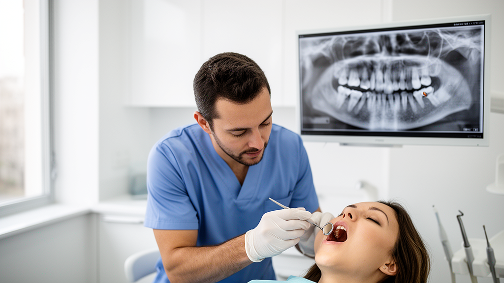
Deep inside the jawbones of children around the world, teeth are quietly under attack. A condition known as pre-eruptive intracoronal resorption, or PEIR, eats away at the dentin of teeth that have not yet broken through the gum line, hollowing out the crown before the tooth has ever been seen by a dentist’s eye. The lesions are invisible to clinical examination and appear only as faint radiolucent shadows on radiographs, typically sitting just beneath the dentinoenamel junction where the protective enamel meets the underlying dentin. Now, a new cross-sectional study from King Abdulaziz University Dental Hospital in Jeddah, Saudi Arabia, has revealed just how easily these hidden defects slip past the professionals trained to catch them, exposing a striking gap between routine diagnostic confidence and the specialized knowledge needed to spot one of pediatric dentistry’s most elusive conditions.
The research, published in Frontiers in Oral Health, set out to measure how well dental practitioners at varying stages of training could detect, diagnose, and manage PEIR. A team led by Amani A. Al Tuwirqi recruited 125 participants from a pool of 163 invited dentists and interns, achieving a response rate of 76.7 percent. The participants were divided into four groups based on educational level: dental interns, general dentists, postgraduate pediatric dentistry residents, and postgraduate residents from other dental specialties. Each participant was then asked to examine a series of eight orthopantomograms, or panoramic radiographs, under standardized viewing conditions in a dimly lit room with a fixed screen distance of 50 to 75 centimeters. The radiographs included one normal image, one case of tooth impaction associated with a dentigerous cyst, one case of odontodysplasia, and five cases of PEIR with varying degrees of severity.
The results paint a picture of practitioners who are highly competent when faced with obvious pathology but strikingly vulnerable when confronted with the subtle radiographic signature of PEIR. Nearly all participants, 98.4 percent, correctly detected the impacted tooth in the second case, and 85.6 percent identified the odontodysplasia affecting multiple teeth. Yet when it came to the moderate PEIR lesion hidden in the third case, only 8.8 percent of participants managed to identify the affected tooth. Even when participants were explicitly told that a radiograph contained an abnormality and asked to locate it, just 16.8 percent correctly identified the PEIR-affected tooth in the second scenario. These numbers suggest that the problem is not simply one of inattention but of a fundamental gap in the visual pattern recognition that experienced radiologists develop through repeated exposure to a condition.
Interestingly, the study found that detection improved dramatically as lesion severity increased. When participants were informed that the cases presented PEIR lesions, correct identification rates rose to 63.2 percent for mild lesions, 54.4 percent for moderate lesions, and 84.8 percent for severe lesions. The researchers attribute this pattern to the physical characteristics of the lesions themselves: larger PEIR defects produce more extensive radiolucency and greater loss of dentinal structure, making them more conspicuous on two-dimensional panoramic images. Mild lesions, by contrast, are often so subtle that they can be easily overlooked during routine radiographic assessment, particularly when the clinician is not actively searching for them.
But here is where the story takes a troubling turn. While participants became better at spotting PEIR as severity increased, their ability to choose the correct management strategy actually declined. For mild lesions, 77.2 percent of participants who identified the affected tooth correctly selected follow-up until eruption as the appropriate approach. For moderate lesions, that figure dropped to 58.8 percent. And for severe lesions, only 32.1 percent correctly chose surgical intervention and restoration before eruption. This inverse relationship between detection confidence and management accuracy suggests that practitioners may recognize that something is wrong but lack the conceptual framework to decide what to do about it, a gap that becomes more dangerous as the lesion becomes more advanced.
The knowledge questionnaire results were equally revealing. Only 24.8 percent of participants had ever heard of PEIR, and a mere 6.4 percent had encountered a PEIR lesion in their own clinical practice. When asked about the underlying cause of PEIR lesions, just 8.8 percent correctly identified resorption as the mechanism. The remaining participants were divided between those who thought it was a localized developmental anomaly, those who attributed it to an infective primary tooth, those who blamed caries, those who suspected fluoride exposure, and those who simply did not know. Despite this widespread unfamiliarity, 98.4 percent of participants acknowledged that dental practitioners should know about PEIR, suggesting that the gap is one of education rather than motivation.
The study also identified clear patterns related to training and experience. Pediatric dentistry residents achieved significantly higher diagnostic scores than dental interns, with a mean diagnosis score of 2.13 compared to 1.72. They also scored significantly higher on PEIR management decisions, with a mean of 2.60 compared to 1.33 for interns and 1.47 for residents from other specialties. Similarly, participants with five or more years of clinical experience outperformed those with only one year of experience across multiple measures, including overall diagnostic scores and PEIR-specific management decisions. These findings align with previous research showing that radiographic interpretation skills improve with specialized training and accumulated clinical exposure, though the authors caution that some subgroup analyses involved relatively small sample sizes and should be interpreted as exploratory.
The biological basis of PEIR adds another layer of complexity to the diagnostic challenge. Histological analyses have revealed that these lesions are characterized by scalloped margins and the presence of chronic inflammatory cells, osteoclasts, macrophages, and multinucleated giant cells originating from the surrounding bone. The condition arises when the protective enamel epithelium is disrupted, allowing resorptive cells to invade the coronal dentin of the unerupted tooth. The prevalence of PEIR in the permanent dentition ranges from 0.6 percent to 27.3 percent depending on the population studied, though its prevalence in primary teeth remains unknown because radiographs are infrequently obtained for unerupted primary teeth. Early diagnosis is crucial because it allows clinicians to monitor lesion progression and intervene before the resorption reaches the pulp or necessitates extraction.
Management of PEIR requires a nuanced understanding of lesion behavior and eruption timing. Conservative monitoring every six to twelve months is often appropriate for small, non-progressive lesions or teeth approaching eruption, with restorative treatment deferred until the tooth can be safely accessed. For symptomatic or rapidly progressing lesions, surgical exposure and curettage may be necessary, ideally without the use of a handpiece to minimize the risk of pulp exposure. When pulp exposure does occur, direct pulp capping with materials such as mineral trioxide aggregate or Biodentine is recommended over traditional calcium hydroxide due to their superior sealing properties. In extensive cases where the lesion is unmanageable, extraction may be the only viable option to preserve overall dental health.
The findings from this study carry significant implications for dental education and clinical practice. The authors argue that undergraduate dental curricula may provide insufficient exposure to uncommon developmental anomalies like PEIR, contributing to reduced awareness and diagnostic confidence among new practitioners. They call for greater emphasis on case-based learning, radiographic interpretation training, and continuing professional education to improve practitioners’ ability to detect and manage PEIR lesions at an early stage. Given that delayed diagnosis can allow lesion progression and increase the risk of pulpal involvement, the stakes extend beyond academic interest to real clinical outcomes for young patients whose developing teeth may be silently compromised. The study also highlights the potential value of three-dimensional imaging modalities such as cone-beam computed tomography, which the authors suggest may offer greater diagnostic sensitivity than the two-dimensional panoramic radiographs used in this research. As awareness of PEIR grows within the dental community, the hope is that practitioners will begin to look more carefully at the unerupted teeth visible on every panoramic radiograph, recognizing that what appears to be a normal developing dentition may sometimes conceal a lesion that is quietly reshaping the tooth from within.
Subject of Research: Dental practitioners' knowledge and diagnostic ability regarding pre-eruptive intracoronal resorption
Article Title: Assessing knowledge and awareness of dental practitioners in detecting, evaluating, and managing pre-eruptive intracoronal resorption: a cross-sectional study
Article References: Al Tuwirqi, A. A., Almashaikhi, J. A., Felemban, O. M., & Elkhodary, H. M. (2026). Assessing knowledge and awareness of dental practitioners in detecting, evaluating, and managing pre-eruptive intracoronal resorption: a cross-sectional study. Frontiers in Oral Health, 7, Article 1880719. https://doi.org/10.3389/froh.2026.1880719
Image Credits: AI Generated
DOI: 10.3389/froh.2026.1880719
Keywords: pre-eruptive intracoronal resorption, PEIR, dental practitioners, radiographic diagnosis, panoramic radiographs, pediatric dentistry, dental education, tooth resorption, Saudi Arabia, cross-sectional study, clinical experience, diagnostic accuracy
Cite Scienmag News
Ophelia Keating. (October 16, 2026). Hidden Tooth Defects Slip Past Dentists: Study Reveals Widespread Blind Spot in Dental Radiology. Scienmag. https://scienmag.com/hidden-tooth-defects-slip-past-dentists-study-reveals-widespread-blind-spot-in-dental-radiology/
Ophelia Keating. "Hidden Tooth Defects Slip Past Dentists: Study Reveals Widespread Blind Spot in Dental Radiology." Scienmag, 16 October 2026, https://scienmag.com/hidden-tooth-defects-slip-past-dentists-study-reveals-widespread-blind-spot-in-dental-radiology/. Accessed 16 October 2026.
Ophelia Keating. "Hidden Tooth Defects Slip Past Dentists: Study Reveals Widespread Blind Spot in Dental Radiology." Scienmag. October 16, 2026. https://scienmag.com/hidden-tooth-defects-slip-past-dentists-study-reveals-widespread-blind-spot-in-dental-radiology/


