


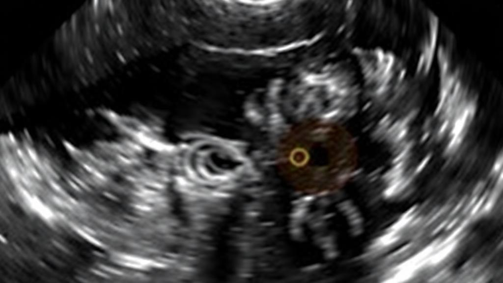
Triple-negative breast cancer has long been regarded as the most visually aggressive form of the disease on imaging scans, typically appearing as an irregular mass with spiky, ill-defined borders. But new research from Türkiye suggests that this aggressive cancer subtype can sometimes masquerade as something far more innocent on ultrasound, a finding that could reshape how radiologists interpret breast scans and prioritize suspicious lesions.
The study, published in BMC Medical Imaging, analyzed 173 invasive breast carcinomas treated at a single Turkish center between 2018 and 2023. Researchers Şahinde Atlanoğlu and Figen Aslan from Kütahya Health Sciences University systematically re-examined mammographic and ultrasonographic images using a standardized reporting lexicon, looking for patterns that might predict which molecular subtype a tumor belonged to before any tissue sample was taken.
What they discovered was unexpected. Two ultrasound features — a circumscribed, smooth-appearing margin and increased blood flow detected on Doppler imaging — were independently associated with the triple-negative phenotype, which lacks estrogen receptors, progesterone receptors, and excess HER2 protein. This subtype is typically the hardest to treat and carries a poorer prognosis than hormone-responsive forms of the disease.
The counterintuitive association between a benign-looking margin and an aggressive cancer challenges conventional radiological wisdom. Most teaching emphasizes that cancers with irregular, spiculated borders are more likely to be high-grade and biologically aggressive. Yet the triple-negative tumors in this cohort frequently presented with well-circumscribed edges, mimicking the appearance of fibroadenomas or other benign lesions.
To ensure their findings were not simply artifacts of subjective interpretation, the researchers had a second blinded radiologist independently re-read 50 of the lesions. Inter-reader agreement was nearly perfect for circumscribed margins and Doppler vascularity, with kappa statistics of 0.90 and 0.96 respectively. This reproducibility is critical, because previous studies linking imaging features to molecular subtypes have often been criticized for inconsistent descriptor use across different observers and institutions.
Not every imaging feature held up to scrutiny. The echo pattern of the lesion — how its internal texture appears relative to surrounding tissue — showed only slight agreement between readers, with a kappa of just 0.19. Isoechogenicity, meaning the lesion appeared similar in brightness to the surrounding breast tissue, was initially associated with triple-negative status but lost its statistical significance once the researchers controlled for margin characteristics in their multivariable model.
The team used Firth penalized logistic regression, a statistical method particularly suited to small sample sizes and rare outcomes, restricting their analysis to variables available before biopsy. After adjusting for confounders, circumscribed margin carried an odds ratio of 3.57 and Doppler vascularity an odds ratio of 3.07 for predicting the triple-negative phenotype. The optimism-corrected area under the curve was 0.71, indicating moderate discriminative ability.
Beyond subtype prediction, the study also examined axillary ultrasound performance for detecting lymph node metastases. The technique proved highly specific at 97.3 percent but only moderately sensitive at 75.8 percent, meaning a normal-appearing axilla on ultrasound does not reliably rule out nodal spread. This has implications for surgical planning, where axillary ultrasound results often influence whether a patient undergoes sentinel node biopsy or more extensive dissection.
Mammographic features, by contrast, showed weaker and less consistent associations. Margin characteristics on mammography differed between subtypes before statistical adjustment but lost significance afterward. Only 52 of the 173 lesions, roughly 30 percent, had mammographic images available for analysis, limiting the statistical power of that component of the study.
The authors are careful to frame their findings as exploratory. The single-center retrospective design, the modest sample size, and the exclusion of patients who received neoadjuvant therapy all limit generalizability. Larger, prospective, multi-center studies with independent image reading will be needed before these imaging signatures can be incorporated into clinical decision-making. Still, the idea that a smooth-bordered, well-vascularized mass on ultrasound might warrant heightened suspicion for triple-negative biology — rather than reassurance — represents a provocative shift in how radiologists might approach the everyday work of breast cancer detection.
The biological explanation for why triple-negative tumors can appear deceptively well-circumscribed on ultrasound remains an active area of investigation. Triple-negative carcinomas are defined by the absence of estrogen receptors, progesterone receptors, and HER2 overexpression, and they disproportionately affect younger women and carriers of BRCA1 mutations. Histologically, many of these tumors grow with a pushing rather than infiltrative border, sometimes accompanied by a prominent lymphocyte infiltrate at the tumor edge. That pushing growth pattern can translate into a smooth, rounded contour on sonography even while the tumor itself behaves aggressively, offering a plausible bridge between the imaging observation and the underlying pathology. The association with Doppler vascularity fits this picture as well, since rapidly proliferating tumors typically recruit new blood vessels, a process that can manifest as increased internal blood flow on color Doppler examination.
The methodological choices in this study deserve attention because they address common weaknesses in the imaging-subtype literature. By re-reading every examination under the fifth edition of the BI-RADS lexicon with a radiologist blinded to pathology results, the authors reduced the risk of circular reasoning, in which readers unconsciously assign descriptors they believe correlate with aggressive biology. The use of Benjamini-Hochberg adjustment across multiple comparisons guards against the false positives that arise when dozens of imaging features are tested simultaneously. Firth penalized logistic regression, meanwhile, is designed for situations where outcome events are sparse and conventional maximum-likelihood estimates can be biased or fail to converge, which is a realistic concern when only about a third of a modest cohort carries the phenotype of interest. Bootstrap internal validation, which produced the optimism-corrected area under the curve of 0.71, provides a more honest estimate of model performance than apparent statistics calculated on the same data used to fit the model.
Even so, an area under the curve of 0.71 falls well short of the discrimination needed for imaging features alone to drive clinical decisions. In practical terms, a circumscribed margin or increased vascularity shifts the probability of triple-negative biology upward but leaves substantial uncertainty. The clinical value of such associations, if confirmed, would more likely lie in combination with other pre-biopsy information, such as patient age, family history, and lesion location, rather than as standalone predictors. It is also worth noting that the analysis set consisted of 173 invasive carcinomas in women, with the lesion rather than the patient as the unit of analysis, and that surrogate subtyping by immunohistochemistry is itself an approximation of the gene-expression-based classifications used in contemporary oncology.
The axillary ultrasound findings carry independent practical weight. A specificity of 97.3 percent means that when ultrasound shows a clearly abnormal node, the likelihood of true metastatic involvement is high, which can prompt needle biopsy and alter surgical planning before the operating room. The sensitivity of 75.8 percent, however, reinforces long-standing guidance that a normal-appearing axilla cannot substitute for nodal staging, since roughly one in four metastatic nodal basins will escape sonographic detection. This asymmetry between ruling in and ruling out disease mirrors the performance of axillary ultrasound reported across broader literature and underscores why sentinel node biopsy remains the standard for surgical staging in most patients.
The mammographic component of the study illustrates how data availability shapes conclusions. With mammograms available for only 52 of the 173 lesions, roughly 30 percent of the analysis set, the power to detect subtype differences was inherently limited, and the loss of significance for mammographic margin after adjustment may reflect either a genuinely weaker association or simply insufficient numbers. In many screening and diagnostic settings mammography is routine, but in this retrospective cohort its absence for most patients likely reflects referral patterns, patient age, or institutional practice, a reminder that single-center imaging studies inherit the biases of the populations they serve.
The reproducibility findings may ultimately prove as consequential as the subtype associations themselves. A kappa of 0.19 for echo pattern, indicating only slight agreement between two blinded readers at the same institution, raises questions about how reliably this descriptor can be used in any predictive model, whether in this study or elsewhere. By contrast, the near-perfect agreement for circumscribed margin and Doppler vascularity suggests these are features radiologists can identify consistently, a prerequisite for any descriptor hoping to survive translation from retrospective research into prospective clinical use. The authors’ decision to report both the promising and the fragile findings in equal detail reflects a growing emphasis in the imaging literature on transparent reporting of measurement reliability alongside associations.
For clinicians interpreting these results, the most defensible takeaway is a heightened index of suspicion rather than a change in biopsy thresholds. A smooth-bordered, hypervascular mass in an appropriate clinical context may merit the same urgency as an obviously malignant-appearing lesion, particularly in younger patients or those with risk factors for triple-negative disease. But the authors themselves emphasize that these associations emerged from an exploratory, single-center cohort and require confirmation in larger, prospective, independently read studies before they can inform guidelines or everyday practice. Until such validation arrives, the study stands as a careful, methodologically disciplined challenge to the assumption that benign-appearing imaging features reliably signal benign biology.
Subject of Research: Associations between breast imaging features and immunohistochemistry-defined molecular subtypes of breast cancer
Article Title: Associations of mammographic and ultrasonographic features with immunohistochemistry-defined surrogate subtypes of breast cancer: a single-center retrospective study
Article References: Atlanoğlu, Ş., & Aslan, F. (2026). Associations of mammographic and ultrasonographic features with immunohistochemistry-defined surrogate subtypes of breast cancer: a single-center retrospective study. BMC Medical Imaging. https://doi.org/10.1186/s12880-026-02953-5
Image Credits: AI Generated
DOI: 10.1186/s12880-026-02953-5
Keywords: breast cancer, ultrasonography, mammography, triple-negative, BI-RADS, immunohistochemistry, surrogate subtype, Doppler vascularity, radiology, cancer imaging, axillary ultrasound, tumor subtypes
Cite Scienmag News
Nathaniel Bowman. (October 16, 2026). Benign-Looking Ultrasound Features May Hint at Aggressive Triple-Negative Breast Cancer. Scienmag. https://scienmag.com/benign-looking-ultrasound-features-may-hint-at-aggressive-triple-negative-breast-cancer/
Nathaniel Bowman. "Benign-Looking Ultrasound Features May Hint at Aggressive Triple-Negative Breast Cancer." Scienmag, 16 October 2026, https://scienmag.com/benign-looking-ultrasound-features-may-hint-at-aggressive-triple-negative-breast-cancer/. Accessed 16 October 2026.
Nathaniel Bowman. "Benign-Looking Ultrasound Features May Hint at Aggressive Triple-Negative Breast Cancer." Scienmag. October 16, 2026. https://scienmag.com/benign-looking-ultrasound-features-may-hint-at-aggressive-triple-negative-breast-cancer/


