


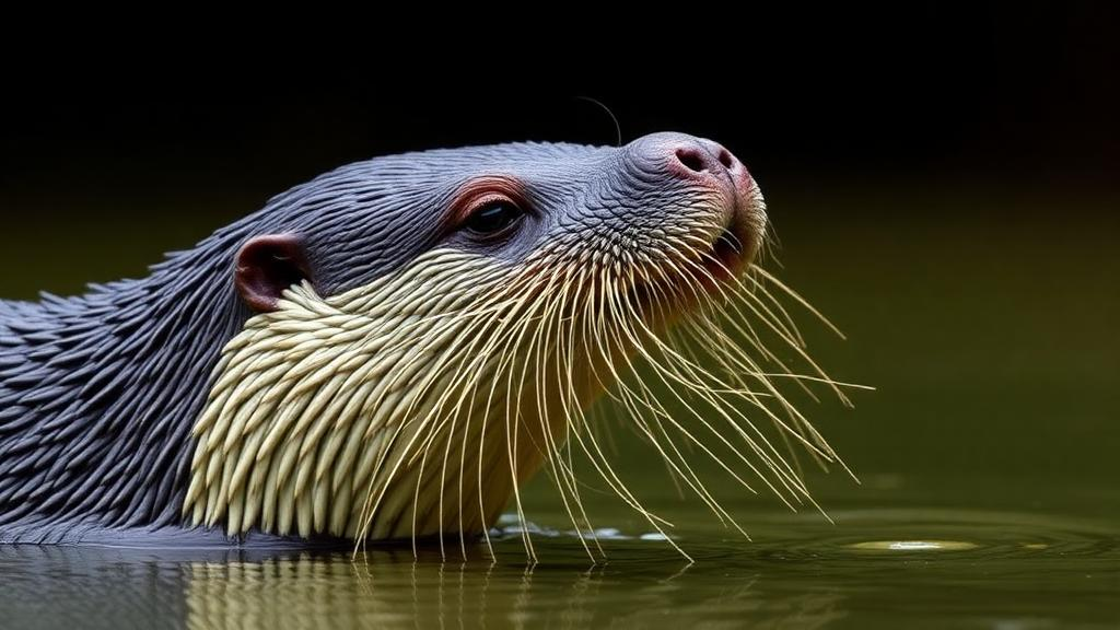
In the winding rivers and oxbow lagoons of Brazil’s Pantanal, the world’s largest otter species is quietly losing its sight. A decade-long study has documented, for the first time, a series of eye disorders and associated systemic illnesses in free-ranging giant otters (Pteronura brasiliensis), the endangered apex predators of South America’s freshwater ecosystems. Using nothing more than high-definition video frames and photographs, wildlife veterinarians identified 12 adult otters with visible ophthalmic abnormalities between December 2009 and October 2019, revealing a potential health crisis that may threaten the long-term survival of local populations in the southern Pantanal.
The findings, published as a case report in Frontiers in Mammal Science, represent the first documented series of ocular disorder cases in wild giant otters. Until now, no published data on eye diseases in this species existed at all, a gap the researchers attribute to the animal’s endangered status, its low population densities, and the extraordinary difficulty of capturing affected individuals from highly social territorial groups. The only published capture method relies on funnel-shaped net traps installed at den entrances, a technique that cannot target specific individuals and depends on narrow environmental windows during the dry season.
What makes this study remarkable is its methodology. Because the otters were never captured or physically restrained, the researchers relied entirely on remote visual documentation. A Sony HDR-CX220 camcorder recording at 1920×1080 full high-definition resolution, supplemented occasionally by a Canon Rebel T6i camera with 70–200 mm L-series lenses, captured footage during routine boat and vehicle surveys. Two wildlife veterinarians then scrutinized the video frames frame by frame, selecting those showing apparent ophthalmic lesions. Certain anatomical features, such as changes in the color of ocular tissues including opacity, can be detected reliably through standard external eye photography, a technique long established in veterinary ophthalmology for examining the ocular surface, eyelid adnexa, and surrounding facial structures when physical examination is impossible.
Each otter was individually identified by its distinctive white throat and chest markings, and the team recorded estimated age, sex, social hierarchy, group composition, and GPS location. Because no species-specific body condition method exists for otters, the researchers adapted a five-point body condition scoring scale used in domestic and wild animals, ranging from 1 for severely emaciated individuals with prominent bones to 5 for obese animals with clearly visible fat deposits. They also scored physical debilitation on a three-level scale based on lethargy and responsiveness to human presence, noted the presence or absence of fresh trauma, and integrated all criteria into an overall health status classification. An individual was deemed clinically unhealthy if any single parameter scored low, ensuring a deliberately conservative assessment.
The results paint a troubling geographic pattern. Ten of the twelve cases, 83.3 percent, occurred in the southern Pantanal, concentrated along the Miranda River, the Negro River, and the lentic pools along the Estrada Parque Pantanal road. Seven otters showed bilateral ocular disorders, two had disease only in the left eye, and three in the right. The earliest records date to December 2009, when two dominant individuals near the Estrada Parque Pantanal were found with swollen, hyperemic mucous membranes in the anus and mouth alongside chronic blepharitis, inflammation of the eyelids, in both eyes. Both animals carried a body condition score of 2, indicating thin body reserves.
The most dramatic sequence unfolded on the Miranda River in 2019. In April and May 2015, a group of three otters had already shown ocular discomfort, with the dominant male so visually impaired that he nearly collided with the researchers’ boat and failed to notice their presence at the group’s campsite. Two of the females in that group had been recorded as clinically healthy just months earlier in November 2014. Then, between August and September 2019, a new group of four otters was observed, three of which developed ophthalmic lesions. One individual showed suspected corneal edema in the left eye with mild third-eyelid protrusion in the right. Another, initially thin with a dorsal cutaneous lesion, deteriorated to a body condition score of 1, struggled to climb the riverbank to its den, and later disappeared entirely. A third maintained better condition but still developed unilateral opacity before declining markedly within days. By late September, only three individuals remained visible, and the last sighting of the group that year came three days later.
Perhaps the most haunting case involved a neighboring group of three otters. One adult, of undetermined sex, developed bilateral ocular opacity suspected to be corneal edema. Throughout August the animal appeared clinically healthy despite the eye findings, but by September the right eye showed conjunctival hyperemia, and by October it exhibited partial paralysis and motor impairment suggestive of near-drowning behavior, unable to swim or maintain buoyancy. Both eyes became more opaque and prominently protruding. Researchers observed the animal for roughly two minutes before it submerged, and despite extensive searching over several hours, neither the otter nor its carcass was ever found. The remaining group members engaged in intense vocalizations and active patrolling, behaviors consistent with searching for a lost companion. The animal was never seen with the group again.
What could be behind these conditions? The researchers propose several candidate etiologies, each grounded in veterinary literature on related mustelids. Toxoplasma gondii, a protozoan parasite shed in the feces of felids, causes significant morbidity and mortality in mustelids; in southern sea otters it was detected in 52 percent of necropsied animals, and clinical signs in otters include corneal edema, uveitis, blindness, ataxia, head tilt, tremors, weight loss, and lethargy. The clinical presentation of the Miranda River otters closely matches this profile, and those groups inhabited areas near a large bridge adjacent to a riverside community, where proximity to humans, domestic animals, and potentially contaminated water or prey could raise exposure risk. Canine adenovirus type 1 offers another possibility: it produces incoordination, generalized ataxia, and bilateral ocular opacity known as the blue eye phenomenon, and it has proven fatal in a Eurasian otter and infectious in sea otters. In the Pantanal, domestic dogs are generally unvaccinated, with rabies often the only vaccine administered.
Environmental contamination adds a further layer of concern. Blindness in Eurasian otters in Britain has been linked to organochlorine insecticides such as dieldrin, which induce hypothyroidism, disrupt vitamin A metabolism, and trigger conjunctivitis, keratitis, and corneal hyperkeratosis. Agricultural land for crops in the Pantanal expanded by approximately 100,000 hectares between 2021 and 2023 according to MapBiomas land-use mapping, intensifying pressure on the wetland. Although no agrochemical residue studies exist for giant otters, elevated mercury concentrations have been reported in fur samples from otters living in the very area where blind individuals were observed along the Miranda River. High mercury is known to cause neurological effects including central blindness, though the authors caution there is no evidence it produces the specific ophthalmic abnormalities seen here. Trauma also plays a role: giant otters engage in extremely aggressive intergroup encounters that leave wounds and scars, and prior research has documented myiasis from such fights leading to death. One otter in this study recovered fully from facial lesions consistent with intraspecific conflict, showing post-inflammatory hyperpigmentation seventy days later, evidence of a self-limiting infection.
The authors emphasize that without capture, clinical examination, and laboratory diagnostics, none of these diagnoses can be confirmed, and the true prevalence and etiology of ocular pathology in giant otters remain unknown. Still, the implications are sobering for a species classified as Endangered by the IUCN since 2000 and Vulnerable on Brazil’s national Red List, one whose range contracted by nearly 40 percent following intensive hunting for the fur trade between the 1940s and 1970s. With no reliable global population estimate and mounting threats from water pollution, fisheries competition, domestic animal diseases, habitat degradation, and climate-driven changes to rainfall and river flow, the accumulation of visible illness in a single river system serves as a warning. The researchers call for improved capture techniques that would allow safe biological sampling, comprehensive clinical and ecotoxicological investigations, and sustainable land-use practices across the Pantanal, before more of these charismatic aquatic predators slip silently into darkness.
Subject of Research: Ophthalmic and systemic disorders documented in free-ranging endangered giant otters in the Brazilian Pantanal
Article Title: Case Report: Ophthalmic and associated systemic disorders in free-ranging giant otters (Pteronura brasiliensis) documented through video frames and photographs
Article References: Soresini, G., Foerster, N., Somma, A. T., da Silva, F. A., Ribas, C., Leuchtenberger, C., & Mourão, G. (2025). Case Report: Ophthalmic and associated systemic disorders in free-ranging giant otters (Pteronura brasiliensis) documented through video frames and photographs. Frontiers in Mammal Science, 4, Article 1634280. https://doi.org/10.3389/fmamm.2025.1634280
Image Credits: AI Generated
DOI: 10.3389/fmamm.2025.1634280
Keywords: giant otter, Pantanal, ophthalmology, wildlife health, Pteronura brasiliensis, Toxoplasma gondii, canine adenovirus, corneal edema, conservation, environmental contamination, Brazil, endangered species
Cite Scienmag News
Margaret Porter. (October 16, 2026). Blindness and Eye Disease Documented in Endangered Giant Otters of the Brazilian Pantanal. Scienmag. https://scienmag.com/blindness-and-eye-disease-documented-in-endangered-giant-otters-of-the-brazilian-pantanal/
Margaret Porter. "Blindness and Eye Disease Documented in Endangered Giant Otters of the Brazilian Pantanal." Scienmag, 16 October 2026, https://scienmag.com/blindness-and-eye-disease-documented-in-endangered-giant-otters-of-the-brazilian-pantanal/. Accessed 16 October 2026.
Margaret Porter. "Blindness and Eye Disease Documented in Endangered Giant Otters of the Brazilian Pantanal." Scienmag. October 16, 2026. https://scienmag.com/blindness-and-eye-disease-documented-in-endangered-giant-otters-of-the-brazilian-pantanal/


