


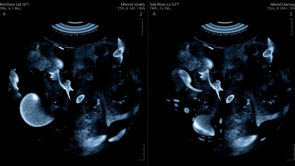
Automated breast ultrasound, or ABUS, has become an increasingly important screening tool because it scans the entire breast in a standardized, operator-independent fashion, reducing the missed-diagnosis problem that plagues handheld ultrasound in dense breast tissue. But the technology carries a hidden vulnerability that has received surprisingly little attention: the gain setting. Gain controls how strongly the returning echo signals are amplified, and it directly determines the brightness and contrast of every image the machine produces. If the gain is set too low, subtle lesions can fade into the background; if it is set too high, tissue texture becomes washed out and boundaries blur. A new study published in BMC Medical Imaging presents a deep learning system that can automatically judge whether an ABUS image was acquired with the right gain, and it does so with an accuracy that rivals careful human review.
The research, led by Mengxia Li, Wenbing Lv, Tongxing Pu, and colleagues at Yunnan University together with clinicians at Yunnan Cancer Hospital, addresses a problem that is easy to overlook. In conventional handheld ultrasound, the sonographer constantly tweaks the gain knob while scanning, drawing on years of experience to keep the image diagnostically useful. ABUS removes much of that human judgment from the acquisition process, which is precisely its strength for reproducibility, but it also means that a poorly chosen gain setting can propagate through an entire automated scan volume before anyone notices. The result is intra- and inter-operator variability that undermines the very standardization ABUS was designed to deliver.
To tackle this, the team assembled what is by any measure a large-scale resource: 31,628 ABUS images collected at Yunnan Cancer Hospital, each labeled by clinical experts into one of five gain quality categories: Insufficient, Low, Optimal, High, and Excessive. That granularity matters. A simple binary judgment of acceptable versus unacceptable would be far easier to learn, but it would also be far less useful in the clinic, where the difference between slightly low and clearly insufficient gain determines whether the image should be re-acquired, adjusted in post-processing, or simply flagged for radiologist awareness. The five-level scheme forces the network to learn a genuinely nuanced visual vocabulary of what good and bad gain look like in real breast tissue.
The architecture they built, named FAMSA-Net for Frequency-Aware Multi-Scale Spatial Attention Network, is built around a simple insight: the fingerprints of incorrect gain are distributed across both spatial scales and frequency bands, and a network that looks for them in only one place will miss them. At its core are gain-discriminative residual blocks, specialized feature extractors designed to respond to the brightness and texture signatures that distinguish one gain level from another. These feed into a Multi-Scale Gain Fusion module, or MSGF, which combines information captured at multiple feature resolutions, so that both fine-grained speckle patterns and coarse tissue-level brightness cues contribute to the final judgment.
Two further components sharpen the system’s perception. The first is Frequency-Domain Enhancement, or FDE, which operates in the Fourier domain rather than the pixel domain. Gain errors leave characteristic traces in the frequency content of an ultrasound image, for example by suppressing or exaggerating particular bands of spatial frequency that correspond to speckle texture and tissue boundaries, and FDE amplifies those cues so the classifier can exploit them more readily. The second is a Spatial Transformer Network, or STN, which performs an learned spatial alignment of the breast anatomy before classification. Because the position and orientation of breast structures vary from patient to patient and scan to scan, this alignment step ensures that the network compares like with like when it judges whether tissue echogenicity falls within the expected range for a given gain setting.
The final piece is Context-Aware Pooling, or CAP, which replaces the conventional global average pooling used in most classification networks. Global average pooling treats every spatial location in the final feature map equally, which discards the fact that some regions of an ABUS image, such as areas near the nipple or deep tissue boundaries, carry far more diagnostic information about gain quality than others. CAP adaptively aggregates contextual information, weighting the contribution of different regions according to their relevance, and the ablation experiments in the paper suggest this step is what allows the other modules to work together effectively rather than at cross purposes.
The headline result is striking. FAMSA-Net achieved 95.49 percent accuracy on the five-class grading task, outperforming two well-established convolutional backbones, DenseNet121 and EfficientNetB3, by margins of 4.19 and 5.56 percentage points respectively. Those baselines are not weak; they are the standard workhorses of medical image classification, and beating them by such margins on a genuinely difficult, fine-grained task is a meaningful result. Just as important for real-world deployment, FAMSA-Net is efficient: it uses only 16.58 million parameters and requires 4.34 GFLOPs of computation, both substantially lighter than the comparison models. A network that could run on standard clinical hardware without exotic accelerators is far more likely to actually reach the ultrasound suite.
The ablation studies, in which individual components were removed to measure their contribution, reveal something more interesting than a simple list of additive wins. Removing the STN caused the largest single drop, with accuracy falling from 95.49 percent to 83.76 percent, confirming that spatial alignment of breast anatomy is the single most critical ingredient in the pipeline. But in a different configuration using global average pooling, removing FDE or MSGF actually improved accuracy, to 90.77 and 90.01 percent respectively. This suggests that the module contributions are not simply additive; the frequency enhancement and multi-scale fusion only pay off when the downstream pooling mechanism can properly exploit the richer representations they produce. When the team replaced global average pooling with CAP, the full model reached its peak of 95.49 percent, demonstrating that contextual aggregation is the glue that makes the whole architecture cohere.
What makes this work notable beyond its benchmark numbers is the problem it chooses to solve. Most deep learning research in medical imaging focuses on the glamorous end of the pipeline: detecting tumors, segmenting lesions, predicting prognosis. Far less attention goes to the unglamorous question of whether the input image is good enough to support those downstream tasks in the first place. Yet image quality is the foundation on which everything else rests, and a screening program that feeds poorly calibrated images into even the best diagnostic model will produce unreliable results. By automating gain quality grading, the Yunnan team has built what amounts to a quality gate at the front of the ABUS pipeline, one that could flag suboptimal acquisitions in real time and prompt re-scanning before the patient leaves the clinic.
The study was approved by the Ethics Committee of Yunnan Cancer Hospital under approval number YJZ2024-22, with written informed consent obtained from all participants, and the work was supported by the National Natural Science Foundation of China and several Yunnan provincial research programs. The authors see their framework as a step toward standardized, objective image acquisition in ABUS, reducing the subjective variability that currently depends on operator experience. As automated screening programs scale up, particularly in regions where sonographer expertise is unevenly distributed, systems like FAMSA-Net may prove to be the quiet infrastructure that makes population-scale ultrasound screening genuinely reliable, one correctly graded image at a time.
Subject of Research: Deep learning-based gain quality grading in automated breast ultrasound images
Article Title: FAMSA-Net: frequency-aware multi-scale spatial attention network for gain quality grading in automated breast ultrasound images
Article References: FAMSA-Net: frequency-aware multi-scale spatial attention network for gain quality grading in automated breast ultrasound images. (n.d.). https://doi.org/10.1186/s12880-026-02861-8
Image Credits: AI Generated
DOI: 10.1186/s12880-026-02861-8
Keywords: automated breast ultrasound, ABUS, gain quality grading, deep learning, image quality assessment, frequency-domain enhancement, spatial transformer network, multi-scale fusion, medical imaging, breast cancer screening, convolutional neural network, BMC Medical Imaging
Cite Scienmag News
Nathaniel Bowman. (October 16, 2026). AI Grades Ultrasound Image Quality With 95% Accuracy in Breast Cancer Screening. Scienmag. https://scienmag.com/ai-grades-ultrasound-image-quality-with-95-accuracy-in-breast-cancer-screening/
Nathaniel Bowman. "AI Grades Ultrasound Image Quality With 95% Accuracy in Breast Cancer Screening." Scienmag, 16 October 2026, https://scienmag.com/ai-grades-ultrasound-image-quality-with-95-accuracy-in-breast-cancer-screening/. Accessed 16 October 2026.
Nathaniel Bowman. "AI Grades Ultrasound Image Quality With 95% Accuracy in Breast Cancer Screening." Scienmag. October 16, 2026. https://scienmag.com/ai-grades-ultrasound-image-quality-with-95-accuracy-in-breast-cancer-screening/


