


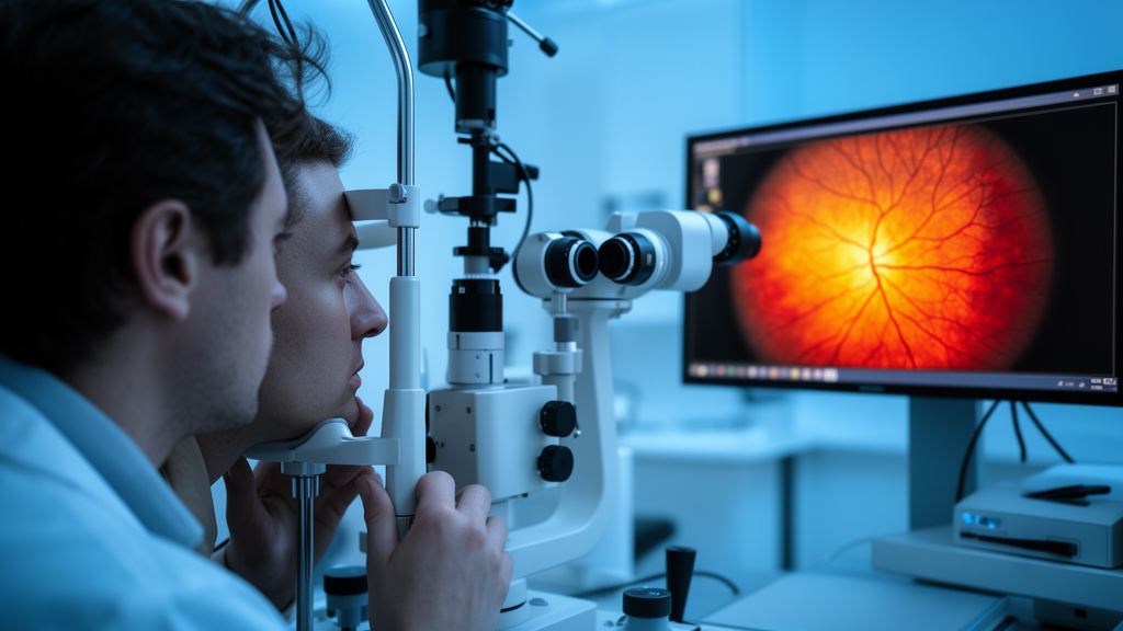
The human eye has long been called a window to the soul, but a new review suggests it may also be a window to hidden cancer. Researchers from the University Clinical Center of Vojvodina and the University of Novi Sad in Serbia have published a comprehensive narrative review in Frontiers in Ophthalmology proposing a structured framework for recognizing when retinal and choroidal abnormalities might signal an underlying systemic malignancy. The work, led by Nikola Denda and colleagues, synthesizes evidence from 52 publications spanning more than two decades and organizes malignancy-related posterior segment findings into five clinically actionable imaging phenotypes that could fundamentally change how ophthalmologists approach unexplained eye findings.
The clinical stakes are considerable. Systemic cancers can reach the eye through several distinct mechanisms: direct metastatic spread through the bloodstream, immune-mediated paraneoplastic attacks on retinal tissue, infiltration by blood or lymphatic cancers, and toxic side effects of anticancer treatments themselves. The choroid, the vascular layer supplying the outer retina, is particularly vulnerable to hematogenous tumor dissemination because of its rich blood flow. Yet the same imaging findings that suggest malignancy can also appear in inflammatory, infectious, vascular, or degenerative disorders, which is precisely why the authors emphasize that imaging alone rarely establishes a diagnosis. Instead, its principal value lies in raising or lowering suspicion, prioritizing competing diagnoses, and guiding the next appropriate investigation.
The review’s central contribution is the phenotype-to-confirmation framework, which organizes suspicious findings into five categories. The first is the exudative choroidal mass phenotype, characterized by an amelanotic or creamy-yellow choroidal lesion accompanied by subretinal fluid, disturbance of the retinal pigment epithelium, and an altered choroidal contour on optical coherence tomography. This combination should raise suspicion for choroidal metastasis, particularly in older patients or those with known cancer. However, the authors caution that amelanotic melanoma, choroidal hemangioma, inflammatory granuloma, and posterior scleritis remain important alternatives that must be excluded before malignancy is presumed.
The second phenotype involves RPE-choroidal remodeling, marked by multifocal changes in the retinal pigment epithelium, abnormal fundus autofluorescence, subretinal fluid, choroidal thickening, and bilateral or diffuse involvement. This pattern may indicate paraneoplastic choroidopathies, most notably bilateral diffuse uveal melanocytic proliferation, a rare syndrome in which ocular changes can precede detection of an occult carcinoma. In BDUMP, highly suggestive findings include multiple round or oval red patches at the level of the RPE, a characteristic patchy or giraffe-like autofluorescence pattern, and progressive outer-retinal disruption. The differential diagnosis here is broad, encompassing central serous chorioretinopathy, Vogt-Koyanagi-Harada disease, posterior scleritis, and drug-related changes, making pattern recognition and longitudinal follow-up essential.
The third category, the vitreoretinal infiltrative phenotype, may be the most clinically urgent because it frequently masquerades as uveitis. Suspicion for vitreoretinal lymphoma increases in older patients with persistent or recurrent intermediate or posterior uveitis, especially when large vitreous cells form sheets or clumps, vitreous haze appears limited relative to the cellular burden, cystoid macular edema is absent or modest, or OCT reveals hyperreflective deposits in the outer retina or sub-RPE space. A transient or incomplete response to corticosteroids does not exclude lymphoma. When suspicion remains high, the authors recommend coordinated diagnostic vitrectomy with prompt processing for cytology, flow cytometry, interleukin-10 and interleukin-6 measurement, immunoglobulin gene rearrangement analysis, and MYD88 L265P mutation testing. Critically, they note that systemic corticosteroids should be withheld before sampling when clinically safe, because prior treatment can reduce cellular yield, and that a single negative test never excludes lymphoma when the clinical course remains suspicious.
The fourth phenotype, diffuse neuroretinal dysfunction, shifts the diagnostic emphasis from structure to function. Patients may experience photopsia, nyctalopia, and visual field loss that appear wildly disproportionate to what the fundus examination shows. Cancer-associated retinopathy and melanoma-associated retinopathy are the principal paraneoplastic culprits, and electroretinography plays a central role in characterization. Cancer-associated retinopathy typically produces generalized rod-cone or cone-rod dysfunction, while melanoma-associated retinopathy classically yields an electronegative ERG pattern, with a relatively preserved a-wave and reduced b-wave, indicating predominantly bipolar-cell dysfunction. The authors stress that antiretinal antibody testing provides only supportive evidence and should never be treated as a standalone confirmatory test, since antibody positivity is not disease-specific and results vary between laboratories.
The fifth category, the treatment-imprint phenotype, addresses a growing challenge in modern oncology: distinguishing therapy-related ocular toxicity from disease progression. Different drug classes produce recognizable but overlapping patterns. MEK inhibitors may cause bilateral, multifocal, shallow subretinal fluid with limited leakage on fluorescein angiography. Immune checkpoint inhibitors can trigger uveitis, retinal vasculitis, or even a VKH-like phenotype with serous retinal detachment. Radiation therapy produces a delayed microangiopathy with hemorrhages, cotton-wool spots, and macular edema. The authors emphasize that a temporal relationship supports but does not prove causality, and that new choroidal or retinal lesions during treatment should never be automatically attributed to toxicity, since metastatic progression may occur during therapy. Decisions about modifying systemic treatment must be made jointly by ophthalmologist and oncologist.
Beyond the five phenotypes, the review offers a sober assessment of artificial intelligence in this domain. While interpretable multimodal models have demonstrated proof of concept in distinguishing choroidal tumor categories, the authors argue that AI should currently be regarded as a research tool rather than an independent diagnostic system for malignancy-related eye disease. The most plausible near-term role is longitudinal comparison rather than single-image classification: tracking lesion dimensions, subretinal fluid, choroidal thickness, and outer-retinal integrity over time. The obstacles are substantial, including small and imbalanced datasets, inconsistent diagnostic reference standards, variable imaging protocols, and device-related domain shift. Current ophthalmic AI evidence remains far stronger in high-volume diseases such as diabetic retinopathy than in these rare and heterogeneous malignancy-related disorders.
The authors are appropriately candid about their own limitations. The narrative design did not include formal risk-of-bias assessment or quantitative evidence grading, and much of the available evidence derives from retrospective series, small cohorts, and diagnostically striking case reports that may overestimate the apparent specificity of individual imaging patterns. The five phenotypes were developed as an interpretive clinical model and have not been prospectively validated; overlap between them is expected, particularly when metastatic, immune-mediated, vascular, and treatment-related mechanisms coexist in the same patient. The framework should therefore be regarded as a structured aid to clinical suspicion and diagnostic escalation rather than a classification system with established sensitivity, specificity, or predictive value.
Nevertheless, the practical message for clinicians and patients is clear: certain findings should lower the threshold for escalation. A new choroidal mass in an older patient, bilateral multifocal abnormalities unexplained by common retinal disease, recurrent or corticosteroid-resistant inflammation, rapid visual deterioration disproportionate to fundus findings, and new posterior segment abnormalities during anticancer therapy all warrant timely exclusion of mimickers and selection of the most appropriate confirmatory step. The authors call for multicenter onco-ophthalmic registries linking ocular findings with cancer type, treatment exposure, and molecular confirmation, which would allow prospective validation of the phenotypes and establish standardized reporting criteria. Until then, the framework offers something genuinely valuable: a disciplined way to convert an atypical eye examination into the right question, asked at the right time, of the right specialist. In a disease landscape where earlier detection can alter trajectories, the retina may prove to be one of medicine’s most underused diagnostic surfaces.
Subject of Research: Posterior segment imaging patterns as clues to systemic malignancy
Article Title: Posterior segment imaging clues to systemic malignancy: a phenotype-to-confirmation narrative review
Article References: Denda, N., Davidović Terzić, S., Maletin, N., Veljkov, K., Rasković, A., Jovančević, L., & Babović, S. (2026). Posterior segment imaging clues to systemic malignancy: a phenotype-to-confirmation narrative review. Frontiers in Ophthalmology, 6, Article 1917738. https://doi.org/10.3389/fopht.2026.1917738
Image Credits: AI Generated
DOI: 10.3389/fopht.2026.1917738
Keywords: choroidal metastasis, multimodal imaging, posterior segment, systemic malignancy, vitreoretinal lymphoma, paraneoplastic retinopathy, optical coherence tomography, BDUMP, anticancer therapy toxicity, electroretinography, artificial intelligence, onco-ophthalmology
Cite Scienmag News
Nathaniel Bowman. (October 16, 2026). Eye Scans May Reveal Hidden Cancers: Five Retinal Warning Patterns Identified. Scienmag. https://scienmag.com/eye-scans-may-reveal-hidden-cancers-five-retinal-warning-patterns-identified/
Nathaniel Bowman. "Eye Scans May Reveal Hidden Cancers: Five Retinal Warning Patterns Identified." Scienmag, 16 October 2026, https://scienmag.com/eye-scans-may-reveal-hidden-cancers-five-retinal-warning-patterns-identified/. Accessed 16 October 2026.
Nathaniel Bowman. "Eye Scans May Reveal Hidden Cancers: Five Retinal Warning Patterns Identified." Scienmag. October 16, 2026. https://scienmag.com/eye-scans-may-reveal-hidden-cancers-five-retinal-warning-patterns-identified/


