


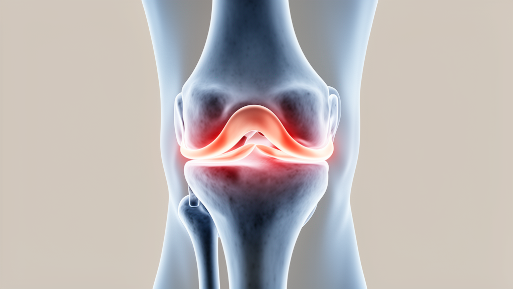
The meniscus is one of the most quietly heroic structures in the human body. Two wedges of tough, rubbery fibrocartilage sit between the thigh bone and the shin bone in each knee, and every step a person takes depends on them. Their secret weapon is an architecture of circumferential collagen fibers that acts like a suspension bridge: when the body’s weight presses down, these hoop fibers convert crushing axial loads into tensile hoop stresses, spreading force across the joint and shielding the cartilage from destruction. When those fibers are severed, the protective machinery fails. A new study published in Frontiers in Bioengineering and Biotechnology has now shown, in striking dynamic detail, how the direction of a person’s leg alignment can either rescue a torn medial meniscus from further harm or push it steadily toward catastrophic failure during ordinary walking.
The research, led by Chengyue Yu, Shuming Cao and colleagues at Tianjin Hospital of Tianjin University together with collaborators in Shandong, Jilin, and Hebei, focuses on a particularly devastating injury: the radial tear of the medial meniscus posterior root, abbreviated MMPRRT. Medial meniscus posterior root tears account for roughly 21.5 percent of all medial meniscus tears, and the radial pattern at the posterior root is the most common subtype of these injuries. The root is the anchoring point where the meniscus fastens to the tibia, and a radial tear there is functionally equivalent to cutting the moorings of the suspension bridge. Hoop stress transmission collapses, local contact pressures surge, cartilage wears faster, and the joint slides toward osteoarthritis. Critically, the authors note, the occurrence of these tears is closely tied to dynamic loading during movement rather than any single static posture.
Most previous biomechanical studies of alignment and meniscal loading have relied on static or quasi-static experiments, capturing only an instantaneous mechanical snapshot at a fixed flexion angle. The Chinese-led team argued that this approach misses precisely the information that matters most: how contact pressure pulses through the joint across a full gait cycle, how the tear gap rhythmically opens and closes under cyclic loading, and how meniscal extrusion, the outward bulging of the meniscus past the edge of the tibial plateau, changes phase by phase. To capture these effects, the researchers built a sophisticated dynamic finite element model of the knee, a computer simulation detailed enough to reproduce the joint’s behavior second by second during a complete walking cycle, and used it to interrogate how shifts in the lower limb’s mechanical axis alter the mechanical environment of the torn root.
The anatomical foundation of the model came from a single healthy male volunteer whose neutral alignment was verified on full-weight-bearing radiographs, with a hip-knee-ankle angle of just 0.44 degrees. High-resolution MRI at one-millimeter slice thickness supplied the soft tissues, including the menisci, articular cartilage, and the major ligaments from the anterior and posterior cruciate ligaments to the collateral ligaments, while CT scans reconstructed the femur, tibia, fibula, and patella. After segmentation, smoothing, and assembly, the resulting model was meshed in Abaqus with more than 773,000 tetrahedral elements, and mesh convergence tests confirmed that predicted tibial cartilage stress varied by less than 2 percent across mesh densities. Cartilage was modeled as a nearly incompressible hyperelastic material using the Yeoh formulation, the menisci as transversely isotropic materials with hoop stiffness far exceeding axial stiffness, and ligaments as nonlinear Neo-Hookean structures.
The study’s methodological centerpiece was a clever way of simulating malalignment without rebuilding the knee. Rather than geometrically bending the femur or tibia to create a varus or valgus deformity, which would confound results with anatomical changes and altered ligament tension, the team simply translated the femoral load application point sideways. Each degree of equivalent varus corresponded to a medial translation of about 3.28 millimeters on a tibial plateau measuring 65.60 millimeters wide. Six conditions were tested on identical geometry: medial shifts equivalent to 3, 6, and 9 degrees, and lateral shifts of 3 and 6 degrees, alongside neutral alignment. Dynamic loading followed the ISO-14243-3 standard gait waveform, the same protocol used for wear-testing knee prostheses, with energy monitoring confirming that inertial artifacts stayed below 5 percent of internal energy throughout the simulation.
Before trusting the model, the team validated it against physical reality. A fresh-frozen cadaveric knee from a young male donor, screened arthroscopically and found free of tears or degeneration, was mounted in a universal testing machine and compressed with a static axial load of 1150 newtons. Ultra-low-pressure Fuji Prescale film, sealed in thin polyethylene bags and slipped into the joint space through a posterior capsular incision, recorded the contact footprint. The cadaveric medial compartment contact area measured 567.98 square millimeters against the model’s prediction of 535.51 square millimeters, an error of only 5.72 percent. Additional plausibility checks against published finite element and experimental studies found the predicted contact pressures fell within reasonable reported ranges, supporting the mechanical credibility of the simulation.
The dynamic results revealed a rhythmic, bimodal pattern of stress that static studies cannot see. Across the gait cycle, peak contact pressures in every knee structure spiked twice, at 14 percent and 44 percent of the cycle, corresponding to the weight-acceptance and push-off phases of walking. At neutral alignment and the first peak, an unstable posterior root tear with a length of 5 millimeters drove medial meniscal pressure up to 22.85 megapascals, compared with 17.82 megapascals for a stable 3-millimeter tear and 16.42 megapascals in the intact knee. The tear gap told an equally dramatic story: at 14 percent of the gait cycle, the unstable tear opened 48.75 percent wider than the stable tear, gaping to 3.57 millimeters against 2.40 millimeters. Maximum meniscal extrusion reached 2.69 millimeters at 44 percent of the cycle, a figure consistent with clinical MRI measurements showing extrusion of around 3 millimeters within a month of symptomatic root tears.
When the team introduced mechanical-axis shifts, the consequences were starkly directional. A medial shift, the computerized equivalent of varus bow-legged alignment, elevated medial peak contact pressures in a strikingly linear fashion: at the mid-cycle static load point, every additional degree of equivalent varus added 1.06 megapascals to medial meniscal pressure, 0.79 megapascals to medial tibial cartilage pressure, and 0.42 megapascals to medial femoral cartilage pressure. Medial shift also prolonged the duration of high overall meniscal displacement, keeping the meniscus in a state of abnormal mobility for a larger fraction of each stride, a finding that aligns with clinical evidence that varus alignment is an independent risk factor for medial meniscal extrusion. The tear gap grew as varus increased from 0 to 6 degrees, with the stable tear widening by 15 percent and the unstable tear by 11.5 percent in the first 3-degree increment alone.
The story at the far end of the varus range grew more complicated, revealing nonlinear behavior that only dynamic modeling could expose. Between 6 and 9 degrees of equivalent varus, the predicted tear gap stopped growing and even declined in some phases, while maximum principal stress in the unstable tear model dropped sharply below its value at 3 degrees. The authors speculate that beyond a threshold of extrusion, the remaining circumferential fibers lose their ability to generate effective hoop stress, the meniscus can no longer disperse load, and the mechanical response enters a different regime. Lateral shifts, by contrast, behaved as a mechanical remedy: valgus-equivalent alignment reduced medial peak contact pressures, shrank the tear gap, and shortened the duration of high meniscal displacement, echoing cadaveric findings that a 3-degree varus alignment raises medial compartment pressure by 37.4 percent while valgus unloads it.
The researchers are careful about the limits of their work. Validation relied on one cadaveric specimen under static loading, the geometry came from a single subject, muscle forces were not actively simulated, and the loading-point method represents a load-distribution type of axis shift rather than true anatomical deformity. These constraints mean the absolute pressures should be read cautiously, even though the relative trends across conditions remain reliable because every simulation shared identical geometry, materials, and loading. Still, the study delivers something genuinely new: a time-resolved map of how alignment and tear size jointly sculpt the mechanical fate of a meniscal root tear during walking. If future prospective clinical studies confirm these associations in patients, the implications could reach rehabilitation planning, identification of high-risk gait phases, and decisions about when alignment-correction surgery should accompany meniscal repair, offering a biomechanically grounded strategy for interrupting the vicious cycle that links misaligned knees, torn menisci, and osteoarthritis.
Subject of Research: Dynamic biomechanical effects of lower limb mechanical-axis shift on medial meniscus posterior root radial tears using finite element gait simulation
Article Title: Dynamic effects of lower limb mechanical-axis shift on medial meniscus posterior root radial tear
Article References: Yu, C., Cao, S., Liu, K., Zhang, J., Zhao, W., Duan, X., Liu, B., Wang, L., Liu, Z., Huo, Y., Zhang, T., Xing, G., & Xu, W. (2026). Dynamic effects of lower limb mechanical-axis shift on medial meniscus posterior root radial tear. Frontiers in Bioengineering and Biotechnology, 14, Article 1925268. https://doi.org/10.3389/fbioe.2026.1925268
Image Credits: AI Generated
DOI: 10.3389/fbioe.2026.1925268
Keywords: knee biomechanics, medial meniscus posterior root tear, finite element analysis, mechanical axis, varus alignment, gait cycle, meniscal extrusion, contact pressure, osteoarthritis, hoop stress, cartilage loading, dynamic simulation
Cite Scienmag News
Denise Maddox. (October 16, 2026). Shifting Loads Reveal How Knee Alignment Worsens Hidden Meniscus Tears While Walking. Scienmag. https://scienmag.com/shifting-loads-reveal-how-knee-alignment-worsens-hidden-meniscus-tears-while-walking/
Denise Maddox. "Shifting Loads Reveal How Knee Alignment Worsens Hidden Meniscus Tears While Walking." Scienmag, 16 October 2026, https://scienmag.com/shifting-loads-reveal-how-knee-alignment-worsens-hidden-meniscus-tears-while-walking/. Accessed 16 October 2026.
Denise Maddox. "Shifting Loads Reveal How Knee Alignment Worsens Hidden Meniscus Tears While Walking." Scienmag. October 16, 2026. https://scienmag.com/shifting-loads-reveal-how-knee-alignment-worsens-hidden-meniscus-tears-while-walking/


