


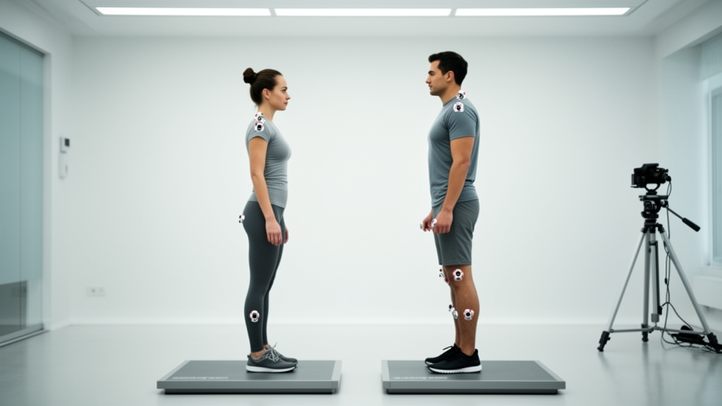
Every spine tells a story, and for the first time, researchers have published the statistical grammar behind it. A team from Fujian University of Traditional Chinese Medicine has compiled reference ranges for 45 upper body posture parameters in 1,859 adults, using a radiation-free imaging technique called DIERS three-dimensional surface topography. The findings, published in Frontiers in Rehabilitation Sciences, reveal something striking: sex, not age, is the dominant force shaping adult posture, with 13 of 45 parameters showing large sex-related effects. The study also uncovered a hidden five-dimensional architecture underlying the posture parameter set, suggesting that the body’s postural system operates as a coordinated whole rather than a collection of independent measurements.
Surface topography works by projecting raster lines onto the back and reconstructing a three-dimensional model from their curvature, a technique known as video raster stereography. Unlike radiography, it involves no ionizing radiation, making it suitable for repeated screening and long-term monitoring. The DIERS system automatically identifies anatomical landmarks, such as the vertebra prominens at the base of the neck and the lumbar dimples on the lower back, and generates quantitative parameters spanning trunk length, spinal curve angles, pelvic obliquity, vertebral rotation, and shoulder alignment. Previous research has demonstrated strong correlations between surface topography parameters and radiographic Cobb angles, with correlation coefficients exceeding 0.8, supporting its role as a complementary screening tool.
The study population comprised 980 men and 879 women with a mean age of 46.9 years, drawn from a health management center database collected between 2014 and 2021. Participants stood on a testing platform while the examiner projected light from above the neck to below the buttocks, capturing a 6-second acquisition. The system then calculated 45 parameters across six measurement domains: length measurements, trunk and pelvis imbalances, locations of postural reference points, spinal curve angles, spinal deviation, and shoulder-related parameters. Reference limits were defined as the 2.5th and 97.5th percentiles, providing population-based benchmarks for clinical interpretation.
Sex-stratified analysis revealed statistically significant differences between men and women in 31 of the 45 parameters, with 13 showing large effect sizes (Cohen’s d ≥ 0.80). These large differences were concentrated in distance measurements, postural reference points, spinal curve angles, and shoulder parameters. For example, mean trunk length VP–DM was 483.53 mm in men versus 445.13 mm in women, a difference of nearly 40 mm with an effect size of 1.40. Shoulder distance SL–SR showed an even larger gap, with men averaging 321.03 mm compared to 292.76 mm in women (Cohen’s d = 1.52). The authors suggest that sex-stratified reference values may be more appropriate for clinical interpretation, though they note that future studies should include height, weight, and BMI to determine whether these differences persist after anthropometric adjustment.
Age-related differences, by contrast, were predominantly trivial to small. Of the 27 parameters showing statistically significant differences across age groups, only three reached medium effect sizes (η² < 0.06). The most notable age-related pattern was an increase in thoracic kyphosis with age: the kyphotic angle ICT–ITL rose from 47.41° in participants aged 18–39 years to 54.94° in those aged ≥60 years, with a medium effect size. The flèche cervicale, a horizontal distance measure reflecting cervical sagittal posture, similarly increased from 55.91 mm to 71.20 mm across age groups. These findings align with a previous systematic review reporting an age-related increase in kyphotic angle, suggesting that sagittal spinal structure may be the domain most sensitive to aging.
The screening classification, based on DIERS-derived criteria for pelvic obliquity, vertebral rotation, and apical vertebral deviation, categorized 35.9% of participants as scoliotic posture, 5.8% as scoliosis risk, and 58.4% as unclassified. The proportion classified as scoliosis risk increased with age, from 4.3% in the youngest group to 8.1% in those aged ≥60 years, though the overall distribution did not differ significantly by sex or age. Notably, differences between screening categories were largely confined to parameters related to the classification criteria themselves, with only four parameters showing medium-to-large effects. Sagittal-plane parameters, such as lordotic angles, showed no significant between-group differences, suggesting that the DIERS screening classification primarily captures coronal and axial alterations rather than sagittal-plane changes.
The most methodologically innovative aspect of the study was the application of exploratory graph analysis (EGA), a network psychometric method borrowed from psychological research. Before EGA, unique variable analysis (UVA) removed 21 redundant parameters, retaining 24 non-redundant ones. EGA then identified a stable five-dimension structure, confirmed in 98.5% of 1,000 bootstrap iterations. The five dimensions were: trunk size and segmental length, coronal deviation and axial rotation, sagittal spinal structure and postural balance, upper-body angular asymmetry, and pelvic asymmetry. This dimensional structure suggests that upper body posture parameters may exhibit coordinated patterns of variation rather than changing independently across individual structures.
Dimension 3, the largest cluster with nine parameters, grouped sagittal spinal structure and postural balance together, implying that thoracic curvature, lumbar curvature, and overall sagittal balance may function as an interrelated multisegmented system. Dimension 2, comprising coronal deviation and axial rotation parameters, corresponds closely to the screening indicators used for scoliosis risk classification, suggesting a potential link between this dimension and scoliosis risk. Dimension 5, containing only pelvic torsion and pelvic obliquity, formed an independent pelvic-level assessment dimension. The authors propose that an individual parameter should not be interpreted in isolation but alongside other parameters within the same dimension, though the clinical validity of this dimension-based interpretation requires further evaluation.
The study’s limitations warrant consideration. The cross-sectional design and single-center health check-up population may not represent the broader adult population. Surface topography measurements are influenced by soft-tissue thickness, subcutaneous fat distribution, and body habits, but anthropometric data such as height and weight were not available, precluding assessment of their confounding effects. The screening categories were defined using DIERS-derived parameters without independent validation against radiographic Cobb angle, and should therefore be interpreted as screening signals rather than diagnostic evidence. Nevertheless, the absence of ionizing radiation makes raster stereography more feasible for frequent monitoring as a complement to radiography, particularly in screening pathways where individuals with suggestive findings could subsequently undergo radiographic assessment for diagnostic confirmation.
These findings provide a quantitative foundation for interpreting surface topography results in health management and scoliosis screening. The pronounced sex-related differences suggest that sex-stratified reference values should be adopted in clinical practice, while the five-dimensional structure indicates that future research should consider coordinated changes across multiple anatomical structures. As larger population datasets become available, machine-learning approaches may further improve the accuracy of spinal structure estimation from surface topography, potentially transforming how posture is assessed and monitored across the life course.
Subject of Research: Reference ranges and dimensional structure of upper body posture parameters in adults: a cross-sectional study
Article Title: Reference ranges and dimensional structure of upper body posture parameters in adults: a cross-sectional study
Article References: Yin, L., Ruan, C., Zhuang, X., Wu, L., Ni, X., Tao, J., & Chen, L. (2026). Reference ranges and dimensional structure of upper body posture parameters in adults: a cross-sectional study. Frontiers in Rehabilitation Sciences, 7, Article 1930857. https://doi.org/10.3389/fresc.2026.1930857
Image Credits: AI Generated
DOI: 10.3389/fresc.2026.1930857
Keywords: Reference, ranges, dimensional, structure, upper, body, posture, parameters, adults, cross-sectional, scientific research
Cite Scienmag News
Beatrice Stafford. (October 16, 2026). Sex, Not Age, Shapes Adult Posture: Landmark Study Maps the Body’s Hidden Blueprint. Scienmag. https://scienmag.com/sex-not-age-shapes-adult-posture-landmark-study-maps-the-bodys-hidden-blueprint/
Beatrice Stafford. "Sex, Not Age, Shapes Adult Posture: Landmark Study Maps the Body’s Hidden Blueprint." Scienmag, 16 October 2026, https://scienmag.com/sex-not-age-shapes-adult-posture-landmark-study-maps-the-bodys-hidden-blueprint/. Accessed 16 October 2026.
Beatrice Stafford. "Sex, Not Age, Shapes Adult Posture: Landmark Study Maps the Body’s Hidden Blueprint." Scienmag. October 16, 2026. https://scienmag.com/sex-not-age-shapes-adult-posture-landmark-study-maps-the-bodys-hidden-blueprint/


