


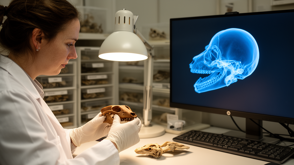
A fragmented skull that sat in a German museum collection for decades, labelled as one of the world’s rarest extinct marsupials, has turned out to be something quite different. Using a handheld 3D scanner and open-source modelling software, researchers digitally reassembled the shattered specimen and discovered it belongs not to the southern pig-footed bandicoot but to a southern long-nosed bandicoot, a common living relative from eastern Australia. The study, published in Natural History Collections and Museomics, demonstrates how virtual reconstruction can rescue the identities of damaged specimens without a single invasive procedure.
The specimen, catalogued as SMNK-MAM 3924 at the State Museum of Natural History Karlsruhe, arrived in 1977 among numerous zoological, palaeontological and geological objects from the collections of the former State Museum of Natural History in Mannheim. Its original label is missing, but a fading ink inscription on the top of the skull reads Chaeropus castanotis, a junior synonym of the southern pig-footed bandicoot (Chaeropus ecaudatus). A second, nearly illegible inscription on the right temporal region shows the number 95 preceded by a short sequence of letters that may read Mai, the German word for May, suggesting either a collection or receipt date of May 1895.
Had the identification been correct, the specimen would have been extraordinarily significant. According to a 2019 review cited in the study, only ten specimens of the southern pig-footed bandicoot are held in Australian museums, with seven others in the Natural History Museum in London and one in the American Museum of Natural History in New York, and nowhere else. The recently described northern pig-footed bandicoot (Chaeropus yirratji) is similarly scarce, known from just fifteen Australian specimens plus single holdings in London, Cambridge and Paris. Most of these specimens were collected during a handful of expeditions between 1838 and 1907, before both species vanished from the semi-arid zones of New South Wales, Victoria, South Australia and Western Australia.
The problem facing curators in Karlsruhe was that the skull had disintegrated into dissociated fragments, most of them fragile, which hampered both morphological evaluation and any attempt at physical reconstruction. Gluing such delicate pieces back together risked further damage and would have permanently altered the specimen. The team instead chose a fully digital route: scan every fragment at high resolution, clean the resulting data, and reassemble the skull on a computer screen.
The scanning was carried out with an Artec Space Spider, a handheld portable 3D scanner whose manufacturer specifications promise accuracy of up to 0.05 millimetres and a resolution of up to 0.1 millimetres. The device weighs just 0.85 kilograms, captures both geometry and surface colour, and operates target-free at 7.5 frames per second on objects as small as five millimetres. Each fragment was placed on a rotating platform to allow a full 360-degree view and scanned from two or three different angles, covering dorsal and ventral or lateral and medial sides. For the two thin, mediolaterally compressed halves of the lower jaw, the researchers borrowed a so-called third hand, a jig normally used in soldering, to hold the elements steady and capture their delicate edges. The scanner’s highest sensitivity setting was used throughout.
Post-processing was performed in ARTEC STUDIO 16 PROFESSIONAL. Unwanted elements such as images of the scanning platform and assorted artefacts were removed manually, and the two to three captures of each fragment were merged into a single 3D image aligned with manually set markers. Incomplete areas of bony surface and residual artefacts were repaired with the software’s lasso selection and fix-holes tools before textures were applied and the files exported in stl, mtl and obj formats. The final assembly took place in Blender 4.1, an open-source 3D modelling package, where published photographs of the skulls of Chaeropus ecaudatus and Chaeropus yirratji served as reference material alongside the actual specimen. The finished renderings were exported as tif files at a resolution of 10,024 by 9,077 pixels.
Six fragments proved sufficient to align into an almost complete skull, with no missing elements added and no parts mirrored. Not everything went smoothly. Attempts to scan the isolated teeth failed entirely, as the scanner’s resolution was not up to the task. The flat, thin dentary halves were prone to bubble-like artefacts on the bone surface that the software could not automatically distinguish from real anatomy, so these had to be removed manually and the areas reconstructed using the surrounding surface as a template. Even at high resolution, slight structural differences between the 3D images and the original fragments required manual correction, though the authors note that none of these adjustments touched any diagnostic characters relevant to species identification. The entire workflow, from scanning through adjustment to virtual reconstruction, took multiple days.
The reconstructed skull told a clear story, and it was not the story on the label. The snout is long and wide, whereas Chaeropus species have short, narrow snouts. There is no accessory palatal fenestra, while Chaeropus always carries a pair of lateral and medial accessory palatal fenestrae. In lateral view the ventral edge of the maxilla is almost straight, rather than showing the deep curvature that elevates the canine and premolars well above the molars in pig-footed bandicoots. The antorbital fossa is weakly developed instead of deep, the zygomatic arch is thin in ventral view rather than thick, and the dentary is very long rather than short, lacking the triangular process at the base of the ascending ramus that expands the dentary laterally in Chaeropus. Features pointing to Perameles include a nasal that terminates well anterior to the anterior margin of the orbit, the presence of the primary foramen ovale, and an alisphenoid tympanic process that is not hypertrophied. The absence of accessory fenestrae, the weak antorbital fossa and the short diastema between the canine and first premolar narrow the field to two species, the southern long-nosed bandicoot (Perameles nasuta) and the northern long-nosed bandicoot (Perameles pallescens). A poorly expanded submasseteric crest on the dentary tipped the balance toward Perameles nasuta, a species that remains common in eastern Australia and even dwells in suburban gardens.
The authors emphasise that the method is remarkably efficient. Without extensive prior training in the hardware and software, scanning took only a few hours, post-processing was completed within a working day, and the virtual reconstruction required a couple of days. One practical limitation emerged: dimensional information could not be transferred from ARTEC STUDIO 16 PROFESSIONAL to Blender through the obj files, so a scale bar had to be adjusted manually using a photograph of the isolated fragments with a scale bar as a template. Dimensional accuracy was then verified by taking real measurements of the left dentary and comparing them with its virtual counterpart. Because the resulting images can be shared online and compared directly with specimens in other museums, virtual reconstruction clearly facilitates identification to species level, a considerable advantage for collections holding damaged or disarticulated material.
How the skull actually reached Mannheim remains an open question. Georg Arnold, who served as curator of the State Museum of Natural History Mannheim from 1876 until his death in 1896, was in contact with the German botanist Ferdinand von Müller in Melbourne, and Arnold’s son Ernst travelled to Australia and collected insects around Melbourne in the 1890s. The researchers consider it not unlikely that either von Müller or Ernst Arnold forwarded the specimen to Mannheim, a hypothesis that fits the faded May 1895 inscription, although no corresponding record exists. The surviving museum catalogues list only very few Australian marsupials and none from Peramelemorphia at all. The latest catalogue entries date from 1877 for mounted mammals and 1901 for skeletons and skulls, yet the museum demonstrably received further specimens at least into the 1930s, including the vast ethnological, palaeontological and zoological collections of Gabriel von Max acquired in 1917. Who misidentified the skull as Chaeropus ecaudatus, and how it entered the collection, therefore remains obscure, a small mystery preserved in fragile bone and fading ink, now at least bearing the right name.
Subject of Research: Virtual 3D reconstruction of a fragmented bandicoot skull to correct its species identification
Article Title: Virtual 3D-reconstruction facilitates identification of fragmented peramelemorph skull
Article References: Specht, J., Manegold, A., & Travouillon, K. J. (2026). Virtual 3D-reconstruction facilitates identification of fragmented peramelemorph skull. Natural History Collections and Museomics, 3, Article e192533. https://doi.org/10.3897/nhcm.3.192533
Image Credits: AI Generated
Keywords: bandicoot, 3D reconstruction, museum collections, Peramelemorphia, Chaeropus, Perameles nasuta, surface scanning, virtual reconstruction, marsupials, taxonomy, natural history, specimen identification
Cite Scienmag News
Margaret Porter. (October 16, 2026). Virtual 3D Reconstruction Reveals Rare Bandicoot Skull Was Mislabelled for Decades. Scienmag. https://scienmag.com/virtual-3d-reconstruction-reveals-rare-bandicoot-skull-was-mislabelled-for-decades/
Margaret Porter. "Virtual 3D Reconstruction Reveals Rare Bandicoot Skull Was Mislabelled for Decades." Scienmag, 16 October 2026, https://scienmag.com/virtual-3d-reconstruction-reveals-rare-bandicoot-skull-was-mislabelled-for-decades/. Accessed 16 October 2026.
Margaret Porter. "Virtual 3D Reconstruction Reveals Rare Bandicoot Skull Was Mislabelled for Decades." Scienmag. October 16, 2026. https://scienmag.com/virtual-3d-reconstruction-reveals-rare-bandicoot-skull-was-mislabelled-for-decades/


