


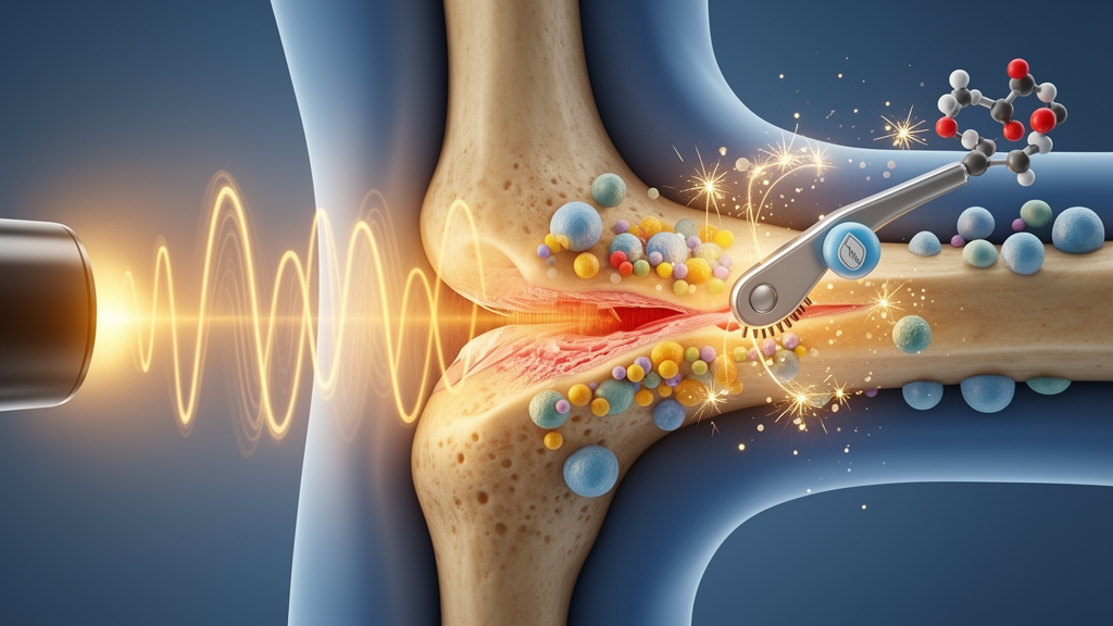
Bone has a remarkable capacity to repair itself, yet large defects, from traumatic fractures to surgical resections, often outpace what natural healing can accomplish. For years, researchers have explored low-intensity pulsed ultrasound, or LIPUS, as a non-invasive way to nudge bone-forming cells into higher gear. Now a team at Capital Medical University in Beijing has mapped a molecular circuit that explains why ultrasound stimulation sometimes falls short, and in doing so has uncovered a promising way to amplify it. Writing in the International Journal of Oral Science, Professor Songlin Wang, Associate Professor Lei Hu and colleagues report that a gene called Id3 acts as a molecular brake on ultrasound-induced bone formation, and that releasing this brake with a purpose-built inhibitory peptide dramatically improved bone-defect healing in mice.
The study begins with a puzzle that has dogged the ultrasound-bone field for some time. LIPUS delivers gentle, pulsed mechanical waves through tissue, and decades of work have shown that these vibrations can activate intracellular signaling cascades that drive mesenchymal stem cells, the raw material of new bone, toward the osteogenic lineage. Yet the technique’s real-world performance in repairing critical-size bone defects has remained inconsistent. That inconsistency suggested to the Beijing team that the body must be deploying internal counterweights, molecular regulators that cap how strongly cells respond to the mechanical stimulus. Identifying those regulators, they reasoned, could reveal new levers for enhancing the therapy.
To find them, the researchers turned to an unbiased discovery strategy. They first performed RNA sequencing on mouse tibiae treated with LIPUS, cataloguing the sweeping changes in gene expression that ultrasound provokes in living bone. From that dataset they compiled a shortlist of 44 transcriptional repressors, proteins whose job is to silence other genes, and screened them with a customized PCR array. One candidate stood out: Id3, short for inhibitor of DNA binding/differentiation 3, showed one of the strongest and most consistent downregulations following ultrasound stimulation, and its known functions pointed directly at processes governing bone. In both mouse bone tissue and mesenchymal stem cells derived from human and mouse bone marrow, Id3 levels fell as LIPUS pushed the cells toward bone formation.
The functional experiments that followed cemented Id3’s role as a genuine negative regulator rather than a bystander. When the researchers forced Id3 overexpression in stem cells, the osteogenic differentiation normally triggered by LIPUS was substantially suppressed, confirming that Id3 actively restrains the bone-forming program. Conversely, stem cells harvested from Id3 knockout mice proliferated faster, differentiated into bone-forming cells more readily, and mineralized their matrices more robustly than cells from normal animals. The knockout mice themselves carried greater bone mass. And when the team delivered extra Id3 to bone tissue using a bone-targeting recombinant adeno-associated virus, designated rAAV9.DSS-Nter-Id3, bone formation dropped. Every manipulation pointed the same direction: less Id3, more bone.
With the brake identified, the next question was what Id3 actually holds in check. Id3 belongs to a family of helix-loop-helix proteins that lack their own DNA-binding domain; instead, they bind and neutralize other transcription factors. The team’s pathway-enrichment analysis of their RNA sequencing data zeroed in on the Bmp4 signaling axis, a canonical driver of osteogenesis. LIPUS treatment, they found, increased Bmp4 expression and set off a downstream cascade: phosphorylation of the signaling proteins Smad1/5, followed by activation of Runx2 and Osx, the master transcription factors that orchestrate bone-cell differentiation. When Id3 was overexpressed, this entire cascade was blunted. And when the researchers pharmacologically blocked BMP signaling with the inhibitor DMH-1, LIPUS could no longer elevate Smad1/5 phosphorylation, and Runx2 and Osx fell back to baseline. The pathway was not merely correlated with the ultrasound response; it was required for it.
The structural logic of the circuit then fell into place. Id3 normally restrains E2A, a basic helix-loop-helix transcription factor capable of binding and activating the Bmp4 gene. Under ordinary conditions, Id3 sequesters E2A and keeps Bmp4 expression in check. When LIPUS drives Id3 levels down, E2A is freed to occupy the Bmp4 promoter more effectively, boosting Bmp4 output and igniting the Smad1/5, Runx2 and Osx cascade that ultimately builds new bone. In effect, ultrasound works partly by loosening a clamp, and the strength of the regenerative response depends on how firmly that clamp is fastened to begin with.
That insight raised an obvious therapeutic question. If Id3 is the brake, could a drug-like molecule release it directly and make ultrasound therapy more powerful? But before the team could design such an intervention, they needed to understand how LIPUS suppresses Id3 in the first place. As Associate Professor Hu noted in the university’s release, knowing that Id3 acted as a brake was only half the story; the upstream mechanism had to be worked out before Id3 could be targeted rationally. A second round of RNA sequencing provided the answer: LIPUS activated calcium-related signaling. Follow-up experiments showed that ultrasound stimulation raised intracellular calcium concentrations, increased the activity of the calcium-dependent phosphatase calcineurin, and triggered the nuclear translocation of NFATC3, a transcription factor that shuttles into the nucleus when calcineurin removes its inhibitory phosphorylations. NFATC3, the team demonstrated, directly suppresses Id3 expression. Prior literature had already established that NFATC3 inhibits Id3, as Dr. Liu explained, allowing the researchers to connect the dots into a complete pathway: CaN/NFATC3/Id3/E2A/Bmp4.
With the full circuit mapped, the team engineered a therapeutic molecule. They created rID3, a peptide designed to disrupt the interaction between Id3 and E2A, functionally mimicking the Id3 downregulation that LIPUS induces but doing so pharmacologically rather than genetically. When rID3 was combined with LIPUS in mouse models of bone defects, the results were striking: osteogenic differentiation was enhanced beyond what ultrasound achieved alone, and bone-defect healing improved measurably. The combination suggests a practical synergy in which mechanical stimulation and molecular brake-release reinforce each other, each compensating for the limitations of the other.
The appeal of such a strategy lies partly in what it avoids. Professor Wang emphasized that if the approach can be materialized clinically, it would offer a convenient, non-invasive route to accelerating bone healing, one that sidesteps the systemic side effects that accompany conventional drug therapies. Ultrasound can be focused precisely on the defect site, and a locally acting peptide that modulates a single protein-protein interaction carries a narrower risk profile than broad-acting osteogenic drugs or anabolic agents. For patients with non-union fractures, spinal fusion failures, or large craniofacial defects, the prospect of a treatment that combines a bedside device with a targeted molecular amplifier is genuinely attractive.
Considerable work remains before the finding reaches the clinic. The current evidence comes from mouse models and cell culture, and the safety, dosing, delivery and long-term consequences of Id3 inhibition in human bone healing have yet to be established. Id3 is also involved in immune-cell development and other biological processes, so systemic interference with the protein would demand careful evaluation. Still, the study delivers something the ultrasound-bone field has lacked: a complete, mechanistically grounded signaling pathway from mechanical stimulus to gene expression, and a concrete molecular target with a proof-of-concept inhibitor already tested in living animals. As sonogenetics matures from an emerging technology into a therapeutic platform, the Id3 brake may well become the reference point against which the next generation of ultrasound-enhanced regenerative therapies is measured.
Subject of Research: The CaN/NFATC3/Id3/E2A/Bmp4 signaling pathway regulating low-intensity pulsed ultrasound-induced bone regeneration
Article Title: Scientists identify Id3 as a molecular brake that could enhance ultrasound-induced bone regeneration
Article References: Scientists identify Id3 as a molecular brake that could enhance ultrasound-induced bone regeneration. (n.d.). Original publication
Image Credits: AI Generated
DOI: Not provided
Keywords: Id3, LIPUS, sonogenetics, bone regeneration, osteogenesis, Bmp4, NFATC3, E2A, mesenchymal stem cells, rID3 peptide, calcineurin signaling, bone-defect healing
Cite Scienmag News
Drew Townsend. (October 16, 2026). Molecular brake discovery points to stronger ultrasound-driven bone healing. Scienmag. https://scienmag.com/molecular-brake-discovery-points-to-stronger-ultrasound-driven-bone-healing/
Drew Townsend. "Molecular brake discovery points to stronger ultrasound-driven bone healing." Scienmag, 16 October 2026, https://scienmag.com/molecular-brake-discovery-points-to-stronger-ultrasound-driven-bone-healing/. Accessed 16 October 2026.
Drew Townsend. "Molecular brake discovery points to stronger ultrasound-driven bone healing." Scienmag. October 16, 2026. https://scienmag.com/molecular-brake-discovery-points-to-stronger-ultrasound-driven-bone-healing/


