


In the summer of his forty-eighth year, a Japanese man arrived at a dermatology clinic in Tokyo complaining of something most people would never think to report: he could no longer tolerate the heat. His body, it turned out, had been quietly losing its ability to sweat for six years. What began as dryness in his palms and soles had crept steadily across his trunk. But the most striking feature of his condition was not where sweating had stopped—it was where it had not. Patches of skin on his left shoulder, right chest, abdomen, and back had become conspicuously dark, and those darkened areas mapped almost perfectly onto the territory where his sweat glands still worked.
The case, reported by researchers at the Institute of Science Tokyo in the Journal of Cutaneous Immunology and Allergy, is a rare and technically fascinating window into the intimate relationship between the autonomic nervous system and the biology of skin pigmentation. The patient had been diagnosed with type 1 diabetes only two years earlier, an acute onset suggested by the absence of high blood glucose in prior checkups. At presentation, his diabetes was poorly controlled, with a hemoglobin A1c of 8.9 percent. Yet his sweating deficit predated his diabetes diagnosis by years, complicating the easy assumption that hyperglycemia alone was to blame.
To understand what was happening, the clinicians turned to classic and modern diagnostic tools in combination. Minor’s method—the iodine-starch test, in which an iodine solution is painted on the skin and dusted with starch so that sweat turns the treated area a deep blue-black—revealed a patchwork of anhidrotic zones: the right shoulder, the left side of the chest and abdomen, and both lower flanks. Infrared thermography corroborated the map. Critically, the hyperpigmented regions corresponded closely to the areas where sweating persisted, an inverse relationship that would become the central puzzle of the case.
Quantitative testing sharpened the picture. In the quantitative sudomotor axon reflex test, acetylcholine is delivered to provoke a local sweating response, allowing sweat output to be measured gravimetrically. On the patient’s back, the left side produced 0.049 milligrams per square centimeter per minute while the right produced 0.159—less than a third of the output. Within the sweating skin itself, Minor’s method revealed heterogeneity, with some zones sweating more vigorously than others, hinting that the surviving nerve supply was unevenly distributed. Brain and spinal magnetic resonance imaging, cardiac sympathetic scintigraphy with iodine-123 meta-iodobenzylguanidine, an active standing test for orthostatic hypotension, and pupillary examination all came back normal, and no sensory or motor neuropathy could be detected. Antibodies associated with autoimmune conditions such as Sjögren’s syndrome were absent. The deficit appeared confined to the sweat apparatus itself.
Skin biopsies from both sides of the back delivered the mechanistic evidence. Eccrine glands in the anhidrotic zones were slightly atrophied compared with those in sweating skin, and immunohistochemical staining for tubulin beta 3—a marker of peripheral nerve fibers—showed a clear reduction in the nerve fibers surrounding sweat glands where sweating had failed. The investigators infer a loss of postganglionic sympathetic fibers, possibly at the level of the sympathetic ganglion, a pattern that echoes segmental anhidrosis syndromes such as Ross syndrome, in which segmental anhidrosis coexists with areflexia and a tonic pupil. That this patient lacked the full Ross triad matters: his condition expands a nosological gray zone between diabetic autonomic neuropathy and the rare idiopathic segmental dysautonomias.
The pigmentation findings were equally revealing. In the skin that still sweated, the basal layer of the epidermis showed greater keratinocyte pigmentation and mild epidermal thickening—52.8 plus or minus 12.1 micrometers versus 31.1 plus or minus 6.7 micrometers in non-sweating skin, excluding the stratum corneum—and dermal collagen fibers were slightly thicker. Crucially, the number of melanocytes was unchanged between the two zones. That single observation shifts the explanatory burden: the difference in coloration must arise from functional changes in melanocyte activity—how much melanin each cell synthesizes and how it deposits pigment—rather than from differences in cell number.
Why would active sweat glands darken the skin above them? The authors propose that the presence or absence of sweating alters the local microenvironment in which melanocytes are embedded. Sweating changes skin hydration, blood flow, temperature, and surface pH, and melanocytes are known to respond to cues from their neighbors. Differences in the state of keratinocytes and fibroblasts between sweating and non-sweating skin could modulate melanocyte function through secreted factors and direct cell-to-cell contacts, thereby shifting melanin production. There is also a possible intensifying effect from the diabetes itself: prior studies have reported lower melanin content in the skin of patients with type 1 diabetes, correlated with glycemic control and the presence of neuropathy. If diabetes drives a background hypopigmentation, the contrast against the relatively hyperpigmented, still-sweating zones would be all the more dramatic—precisely the asymmetrical pattern this patient displayed.
Sweating dysfunction is a well-recognized but underappreciated complication of diabetes. It can manifest as hyperhidrosis, gustatory sweating triggered by food, or anhidrosis and hypohidrosis in variable distributions. The most common pattern, distal hypohidrosis, is attributed to hyperglycemia-induced damage to the long peripheral axons that innervate distal sweat glands, which is why diabetic sweating loss typically begins symmetrically in the feet and hands. This patient breaks the template. His deficit began distally but spread asymmetrically to the trunk years before diabetes onset, and his peripheral neuropathy workup was otherwise clean. The authors argue that the extensive, non-length-dependent distribution of his sweating loss, together with the histological evidence of fiber loss around the glands, distinguishes him from typical diabetic autonomic neuropathy—while acknowledging that because the anhidrotic map does not align neatly with sympathetic segment boundaries, local skin conditions may also modulate sweat gland function.
The clinical stakes are not merely cosmetic. Anhidrosis removes a primary channel of evaporative heat dissipation, and heat intolerance in an anhidrotic patient is a genuine risk factor for heat stroke. With no established method for restoring sweating in segmental or asymmetrical anhidrosis, management is necessarily conservative: the patient was counseled to take frequent breaks in hot environments and during exercise, and to apply wet towels regularly to compensate artificially for his absent evaporative cooling. As global temperatures rise, such practical guidance for patients with thermoregulatory impairment becomes an increasingly visible corner of preventive medicine.
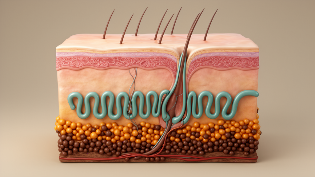
Beyond its clinical message, the case is a reminder that seemingly separate physiological systems are entangled at the scale of single cells. A nerve fiber’s presence around a coiled eccrine gland appears to influence the pigmentation decisions of melanocytes several cell layers above, mediated through the chemistry of sweat, blood flow, and the chatter between keratinocytes and fibroblasts. For scientists studying skin homeostasis, the sweating-dark skin of this one patient offers a natural experiment: two adjacent territories of tissue, genetically identical and innervated by the same body, diverging in both secretory function and color because one lost its autonomic supply. Unraveling which molecular signals link sweat output to melanin synthesis could illuminate not only rare dysautonomias but also everyday questions about why skin darkens, lightens, and ages the way it does.
Subject of Research: Asymmetrical anhidrosis and hyperpigmentation in type 1 diabetes
Article Title: Asymmetrical anhidrosis with hyperpigmentation in a patient with type 1 diabetes
Article References: Fujiwara, M., Iida, T., Oda, A., Takeshita, H., Namiki, T., & Okiyama, N. (2026). Asymmetrical anhidrosis with hyperpigmentation in a patient with type 1 diabetes. Journal of Cutaneous Immunology and Allergy, 9, Article 17110. https://doi.org/10.3389/jcia.2026.17110
Image Credits: AI Generated
Keywords: anhidrosis, hyperpigmentation, type 1 diabetes, sweating, melanocytes, autonomic neuropathy, eccrine glands, Ross syndrome, thermoregulation, Minor's test, skin biopsy, dermatology
Cite Scienmag News
Ophelia Keating. (October 15, 2026). When Sweat Stops, Skin Darkens: A Diabetes Case Reveals a Hidden Link. Scienmag. https://scienmag.com/when-sweat-stops-skin-darkens-a-diabetes-case-reveals-a-hidden-link/
Ophelia Keating. "When Sweat Stops, Skin Darkens: A Diabetes Case Reveals a Hidden Link." Scienmag, 15 October 2026, https://scienmag.com/when-sweat-stops-skin-darkens-a-diabetes-case-reveals-a-hidden-link/. Accessed 15 October 2026.
Ophelia Keating. "When Sweat Stops, Skin Darkens: A Diabetes Case Reveals a Hidden Link." Scienmag. October 15, 2026. https://scienmag.com/when-sweat-stops-skin-darkens-a-diabetes-case-reveals-a-hidden-link/


