


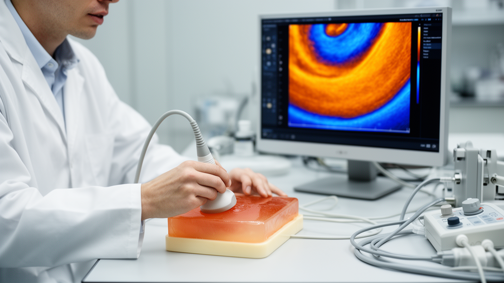
Ultrasound thermal strain imaging (US-TSI) has long promised something remarkable: the ability to peer inside living tissue and read its chemical composition — particularly its fat content — using nothing more than sound waves and a gentle pulse of heat. Now, a team of researchers at the University of Pittsburgh and North Carolina State University has put that promise to one of its most rigorous quantitative tests yet, showing that the technique’s response to temperature and composition can be predicted, simulated, and measured with striking agreement. The study, published in Frontiers in Acoustics, provides the kind of numerical grounding the field has been waiting for.
The core idea behind US-TSI is elegantly simple. When tissue warms even slightly, the speed of sound within it changes. Because the imaging system assumes a fixed sound speed when reconstructing images, this temperature-induced change causes echoes to shift slightly in time, producing what looks like a tiny apparent displacement of the tissue. The spatial gradient of that displacement is called thermal strain. Crucially, water-rich tissues and lipid-rich tissues respond in opposite ways: sound speeds up with temperature in water-bearing material but slows down in fat. That sign difference is what allows US-TSI to distinguish lipid from water — and, in principle, to quantify how much of each is present.
Despite decades of development, most previous studies relied on assumed or literature-based sound speed–temperature relationships and treated thermal strain mostly as a qualitative contrast signal. What has been missing is a unified framework that connects the actual measured acoustic properties of a material, a physics-based mixture model, realistic ultrasound simulations, and controlled experiments — all in one place. That is precisely what the new study delivers, using oil–gelatin phantoms as controlled surrogates for lipid-bearing tissue.
The researchers began by measuring the speed of sound in pure castor oil and in a 5 percent gelatin solution across a temperature range of 24 to 40 degrees Celsius, spanning both room-temperature phantom conditions and physiological temperatures near 37 degrees. The two materials showed opposite temperature dependence: the gelatin solution’s sound speed increased with temperature, while the oil’s decreased. These measured relationships then fed into Wood’s equation, a classical acoustic mixture model that predicts the effective sound speed of a two-component medium from the densities and sound speeds of its constituents.
With the mixture model in hand, the team turned to Field II, a widely used ultrasound simulation platform, to generate synthetic radiofrequency signals for phantoms containing oil mass fractions from 0 to 40 percent. In each simulation, the scatterer positions were held perfectly fixed while only the sound speed changed between a baseline state and a heated state. Beamforming with an assumed constant sound speed of 1,540 meters per second then produced the characteristic apparent displacement, which was tracked using the same Loupas phase-based algorithm applied to the experimental data. The result was an idealized upper-bound estimate of what US-TSI can detect under perfectly controlled conditions.
The experimental side of the study used a Verasonics Vantage 256 system with a focused heating beam delivering 120-cycle pulses over 50 milliseconds at varying transmit voltages from 10 to 40 volts. Imaging was performed with a five-angle compounded plane-wave sequence at 7.8 megahertz and a frame rate of 500 hertz, capturing enough frames for stable displacement estimation. A calibrated thermocouple measured the actual temperature rise, which was cross-validated against a heat-diffusion cooling model fitted to the post-heating temperature decay. That model showed excellent agreement with direct measurements, with a root-mean-square error of just 0.24 degrees Celsius.
The results are striking in their consistency. Thermal strain varied linearly with temperature rise across the tested range of roughly 1 to 4 degrees Celsius, with coefficients of determination reaching 0.975 to 0.996 in simulation and 0.868 to 0.968 in experiment. Even more importantly for tissue characterization, thermal strain varied linearly with oil mass fraction at each fixed temperature rise, with simulation R² values of 0.998 to 0.999 and experimental R² values of 0.925 to 0.993. Across all 35 matched simulation–experiment conditions, the overall root-mean-square error was 0.016 percent strain, corresponding to just 7.8 percent of the total experimental strain range, and the simulated and experimental values correlated at r = 0.95.
The physical mechanism underlying these trends is the opposite temperature dependence of sound speed in the two components. As temperature rises, sound travels faster through the gelatin matrix but slower through the oil droplets, causing the two materials to diverge acoustically. This divergence amplifies the propagation-time mismatch under constant-speed beamforming, producing larger apparent displacements and stronger strain signals. The result is that heating more — pushing the temperature rise higher — effectively amplifies the compositional contrast without changing the underlying linear relationship, a finding that has direct implications for how clinicians might optimize heating protocols in future applications.
The study also quantified the practical detection limits. Using baseline noise measurements from repeated pre-heating acquisitions, the team estimated an empirical strain detection threshold of approximately 1 × 10⁻⁴, or 0.01 percent strain. Low- and intermediate-oil compositions produced strain signals well above this threshold, while compositions near the high-oil zero-crossing — around 35 percent oil mass fraction — approached the noise floor. This means the technique is most reliable for detecting composition-dependent differences rather than pinpointing an exact oil fraction near the point where the strain signal vanishes. The researchers also note that their simulations represent an idealized upper bound, since they excluded motion, signal decorrelation, and electronic noise that would inevitably degrade performance in living tissue.
The implications extend well beyond phantom experiments. Previous work by overlapping members of this team has already demonstrated US-TSI’s potential for detecting lipids in atherosclerotic plaques in rabbits and pigs and for assessing hepatic fat in obese mice. More recently, a millisecond-level transient heating probe achieved approximately 2 degrees Celsius of temperature rise in just 50 milliseconds in a porcine model, opening the door to real-time, clinically compatible thermal strain imaging. By establishing a rigorous quantitative framework that links composition, temperature, and strain through measured material properties, validated mixture modeling, and matched simulation and experiment, this new study provides the numerical foundation that could help translate US-TSI from a promising research tool into a practical method for noninvasive fat quantification in cardiovascular disease, fatty liver disease, and beyond.
Subject of Research: Quantitative validation of ultrasound thermal strain imaging sensitivity for temperature and lipid composition detection
Article Title: Quantitative validation of ultrasound thermal strain imaging sensitivity using simulation and oil–gelatin phantom experiment
Article References: Quantitative validation of ultrasound thermal strain imaging sensitivity using simulation and oil–gelatin phantom experiment. (n.d.). https://doi.org/10.3389/facou.2026.1861076
Image Credits: AI Generated
DOI: 10.3389/facou.2026.1861076
Keywords: ultrasound thermal strain imaging, speed of sound, tissue characterization, lipid detection, Wood's equation, oil–gelatin phantom, Field II simulation, noninvasive thermometry, atherosclerotic plaque, fatty liver, biomedical ultrasound, quantitative imaging
Cite Scienmag News
Carl Richardson. (October 15, 2026). Ultrasound Thermal Strain Imaging Passes a Rigorous Quantitative Sensitivity Test. Scienmag. https://scienmag.com/ultrasound-thermal-strain-imaging-passes-a-rigorous-quantitative-sensitivity-test/
Carl Richardson. "Ultrasound Thermal Strain Imaging Passes a Rigorous Quantitative Sensitivity Test." Scienmag, 15 October 2026, https://scienmag.com/ultrasound-thermal-strain-imaging-passes-a-rigorous-quantitative-sensitivity-test/. Accessed 15 October 2026.
Carl Richardson. "Ultrasound Thermal Strain Imaging Passes a Rigorous Quantitative Sensitivity Test." Scienmag. October 15, 2026. https://scienmag.com/ultrasound-thermal-strain-imaging-passes-a-rigorous-quantitative-sensitivity-test/


