


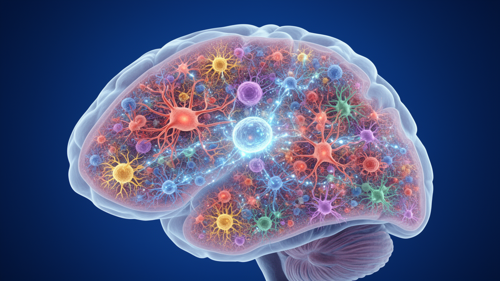
Deep in the forebrain, a structure the size of a walnut in humans quietly decides which of your countless potential movements, thoughts, and impulses gets to become reality. The striatum, the largest nucleus of the basal ganglia, receives a torrent of excitatory signals from nearly every region of cortex, blends them with thalamic traffic and a constant drip of neuromodulators, and converts this convergence into the patterns of activity that ultimately select and execute actions. For decades, textbooks framed this computation around a deceptively simple dichotomy: striatal projection neurons of the direct pathway, which promote movement, versus those of the indirect pathway, which suppress it. A new collection of studies published in Frontiers in Cellular Neuroscience argues that this categorical view has outlived its usefulness, and that the striatum’s true computational power emerges from a far richer interplay of cellular subtypes whose identities, positions, and connections are only now being mapped in molecular detail.
The editorial, authored by Elodie Fino of Aix-Marseille University’s INMED institute, Maxime Assous of Cardiff University, and Marc Vincent Fuccillo of the University of Pennsylvania, gathers eight contributions—four original research articles, two reviews, and two mini-reviews—that together chart the field’s transition from relatively categorical descriptions of striatal cell types toward a multidimensional understanding of cellular identity, spatial organization, connectivity, and function. The central message is provocative in its implications: cellular diversity is not merely a descriptive feature of the striatum. It is a fundamental determinant of how the structure integrates and transforms information, and therefore of how animals move, learn, and respond to reward.
The first theme to emerge from the collection is that neuronal identity is multidimensional, and that the frameworks neuroscientists have relied upon to make sense of striatal activity are more fragile than they appear. Electrophysiological classifications that have guided much of the in vivo literature—phasically active neurons, tonically active neurons, and fast-spiking interneurons—do not necessarily correspond in a straightforward manner to molecularly defined cell types. In a perspective piece, Apicella and colleagues revisit this classical PAN-TAN-FSI framework, emphasizing both its enduring usefulness and its limitations. The categories were built on firing patterns observed in behaving animals, but firing patterns are state-dependent: the same neuron can shift its behavior depending on neuromodulatory context, arousal, and task demands. A cell-type label derived from one dimension of measurement may simply not survive translation into another.
This tension between classification schemes is brought into sharp molecular focus by Naon and colleagues, who provide a detailed translatomic characterization of parvalbumin-expressing interneurons in the dorsal striatum. Using techniques that profile the messenger RNA actively being translated in these cells, the researchers identified a molecular signature that distinguishes dorsal striatal PV interneurons from their counterparts in the nucleus accumbens, the ventral striatal region central to reward and motivation. The finding is striking because it means that a genetically defined population—cells that all express the same marker protein—are not molecularly interchangeable across striatal territories. Parvalbumin interneurons in the dorsal striatum carry a translational identity shaped by their location, and presumably by the developmental history and afferent environment that come with it. Together with the Apicella perspective, the study underscores the importance of integrating molecular identity with electrophysiological properties and circuit function, rather than treating any single feature as sufficient to define a neuronal subtype.
Cellular diversity, the collection further shows, is inseparable from anatomical organization. Muhsinov and colleagues performed a three-dimensional mapping of somatostatin-expressing, parvalbumin-expressing, and tyrosine hydroxylase-expressing interneurons across the mouse dorsal striatum, revealing distinct spatial distributions for each population. This is far more than cartographic trivia. The position of a neuron determines the inputs it receives, the functional territory it belongs to, and the local circuits it can influence. An interneuron sitting at the border between two functional domains may sample afferents from both; one buried deep within a compartment may be wired into an entirely different microcircuit. Spatial organization thus acts as a structural blueprint that constrains which cellular interactions are even possible, and the new mapping suggests that the striatum’s inhibitory interneuron populations are deployed with a precision that reflects this logic.
The spatial dimension becomes particularly consequential in the review by Lemos and colleagues on striatal cholinergic interneurons. Although cholinergic interneurons constitute a relatively small fraction of striatal cells, their influence on striatal networks is vast: they provide the tonically active, acetylcholine-based signaling that has shaped much of the field’s understanding of striatal dynamics, and they sit at the center of the dialogue between acetylcholine and dopamine that governs action selection and reinforcement. Yet their properties and their interactions with dopamine and other neuronal populations differ across striatal territories. A cholinergic interneuron in the dorsal striatum is not functionally equivalent to one in the ventral striatum, even though the two cells would be indistinguishable by conventional genetic markers. Even a genetically defined cell population, the review concludes, may not represent a functionally homogeneous entity—a sobering caveat for any study that treats marker expression as a proxy for uniform function.
If cellular identity and spatial position provide the striatum’s parts list and blueprint, circuit interactions supply the machinery. Striatal activity results from the integration of convergent excitatory inputs and local inhibitory mechanisms, rather than from the activity of individual neuronal classes in isolation, and several contributions illustrate how cell-type-specific interactions are embedded within the striatum’s compartmental architecture. Karube and colleagues added another organizational dimension by showing that pallidostriatal inhibitory projections—the inhibitory traffic arriving from the external segment of the globus pallidus—preferentially target the matrix compartment of the dorsal striatum, including both projection neurons and cholinergic interneurons. The striosome-matrix architecture, a compartmentalization of the striatum that has intrigued anatomists for half a century, thus appears to channel long-range inhibitory inputs in a compartment-specific manner. The findings suggest a concrete route through which globus pallidus externus inputs may shape compartment-specific striatal processing, linking a major basal ganglia output nucleus back to the input structure in a cell-type- and territory-selective fashion.
The functional consequences of such specificity are driven home by Long and Masmanidis, whose mini-review revisits one of the oldest debates in basal ganglia neuroscience: the distinction between dopamine’s roles in reinforcement learning and in movement. Rather than treating these as separate functions of the same neuromodulator, the authors advance a learning primacy hypothesis, arguing that understanding dopamine’s behavioral functions requires resolving how dopaminergic signals act on specific striatal cellular and synaptic substrates across distinct timescales. The proposal has significant implications. If dopamine’s contribution to movement is itself an emergent property of learning-related plasticity at particular synapses on particular cell types, then the search for dopamine’s mechanism must happen at the level of identified cellular substrates, not at the level of global striatal activity. These studies reinforce the view that the functional consequence of a given signal depends critically on the nature of the targeted cells, their location, and how they are embedded within the larger network.
The final theme makes the stakes explicit: understanding cellular subtypes is essential for explaining how striatal circuits become dysfunctional in disease. Nelson and colleagues report developmental alterations in the excitability of striatal projection neurons in the Fmr1–/y mouse model of fragile X syndrome, highlighting the importance of the temporal evolution of cellular dysfunction. The finding suggests that fragile X-related circuit pathology is not a static deficit but a moving target, with projection neuron properties drifting across development in ways that could reshape striatal computation at each stage. In parallel, Thabault and Galvan review the contribution of parvalbumin interneurons to striatal dysfunction in Huntington’s disease and autism spectrum disorder, two conditions with very different etiologies that converge on the same vulnerable cell population. Together, these studies illustrate that pathological changes cannot necessarily be described as a global increase or decrease in striatal activity. Different cell populations may be affected through distinct mechanisms and at different stages, potentially leading to complex and evolving alterations in circuit dynamics that a single activity readout would collapse into misleading simplicity.
Taken together, the Research Topic places cellular heterogeneity at the center of striatal neuroscience. Molecular identity, developmental stage, electrophysiological properties, anatomical position, connectivity, and neuromodulatory sensitivity provide complementary dimensions through which neuronal populations contribute to striatal computation, and no single dimension is sufficient on its own. The timing is apt: recent advances in genetic targeting, transcriptomics, anatomical mapping, and cell-type-specific recording and manipulation now make it possible to examine this diversity across molecular, cellular, circuit, and behavioral scales, transforming questions that were once philosophical into empirically tractable ones. For a structure whose dysfunction lies at the heart of Parkinson’s disease, Huntington’s disease, addiction, and autism, the message is clear. The striatum’s secrets will not be found by averaging across its cells, but by resolving them—one subtype, one compartment, one synapse at a time.
Subject of Research: Neuroscience
Article Title: Editorial: The role of cellular subtypes in striatal activity
Article References: Editorial: The role of cellular subtypes in striatal activity. (n.d.). https://doi.org/10.3389/fncel.2026.2010201
Image Credits: AI Generated
DOI: 10.3389/fncel.2026.2010201
Keywords: Editorial, role, cellular, subtypes, striatal, activity, scientific research
Cite Scienmag News
Drew Townsend. (October 15, 2026). Inside the Striatum: Why Neuronal Subtypes Hold the Key to Action and Disease. Scienmag. https://scienmag.com/inside-the-striatum-why-neuronal-subtypes-hold-the-key-to-action-and-disease/
Drew Townsend. "Inside the Striatum: Why Neuronal Subtypes Hold the Key to Action and Disease." Scienmag, 15 October 2026, https://scienmag.com/inside-the-striatum-why-neuronal-subtypes-hold-the-key-to-action-and-disease/. Accessed 15 October 2026.
Drew Townsend. "Inside the Striatum: Why Neuronal Subtypes Hold the Key to Action and Disease." Scienmag. October 15, 2026. https://scienmag.com/inside-the-striatum-why-neuronal-subtypes-hold-the-key-to-action-and-disease/


