


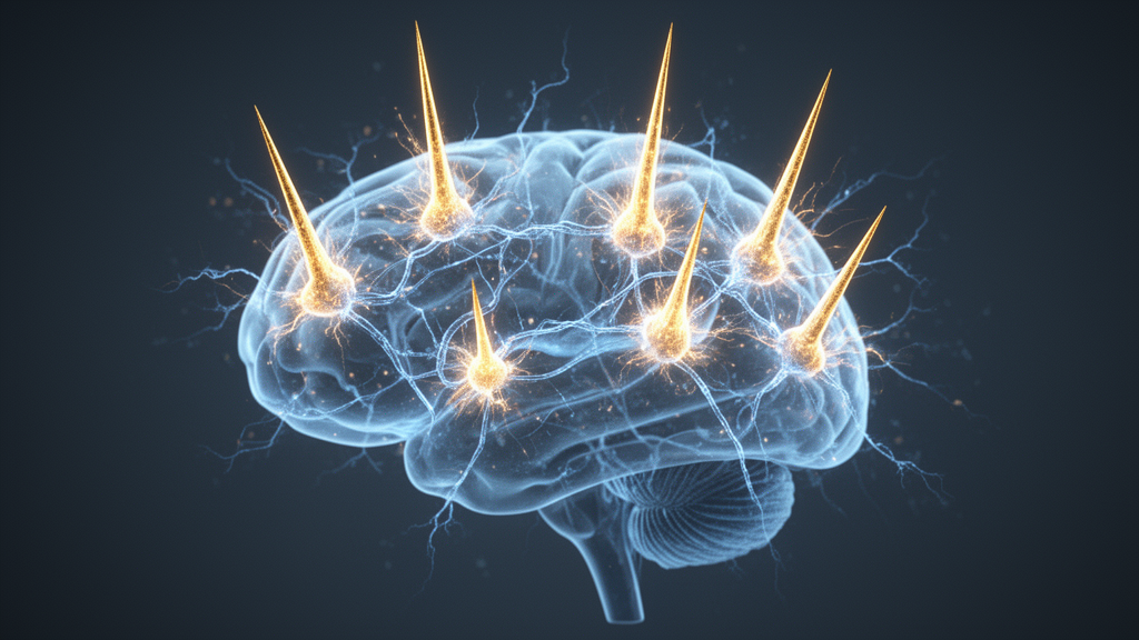
For decades, epilepsy surgery has been built on a deceptively simple idea: find the spot where seizures start, cut it out, and the patient gets better. Yet a substantial number of patients continue to have seizures even after what appears to be a technically successful resection, and roughly one in three people with epilepsy live with a drug-resistant form of the disorder. A new perspective article published in Frontiers in Network Physiology argues that this treatment gap exists because clinicians have been asking the wrong question. Rather than asking where seizures begin, the authors contend, researchers and surgeons should be asking which connected brain structures form the network that generates and sustains them. The article, led by James E. Niemeyer of Weill Cornell Medicine, proposes a framework called network excitability mapping, built on two measurable dimensions: the local excitability of individual brain nodes and the degree to which epileptic activity recruits distributed regions across the brain.
The conceptual shift has deep roots. As early as 2002, the epileptologist Susan Spencer highlighted that the seizure onset zone may not be the true generator of seizures but merely one vulnerable node within a broader network capable of initiating or supporting pathological hyperactivity and synchrony. When seizures persist after resection, they may arise from multiple regions, emerge from latent secondary sites, or persist through network reorganization after surgery. In this view, the key determinant of seizure generation is not only where activity begins, but how pathological excitation propagates through a network, and which nodes amplify or constrain that activity. The problem, the authors argue, is that there are currently few ways to directly measure or map this network-level excitability in either patients or experimental animal models, leaving a gap between mechanistic insights from preclinical neuroscience and the biomarkers used in the clinic.
Computational neuroscience has already embraced the network view of epilepsy. Graph theory, which represents the brain as a set of nodes joined by connections of varying strength, has allowed researchers to quantify properties such as how central a given node is and how readily activity travels between regions, from the scale of single neurons to the whole brain. This approach can identify highly connected hub nodes that exert disproportionate influence over how seizures spread, and when applied to human epilepsy, such network measures have predicted surgical and neurostimulation outcomes more accurately than anatomical localization of the seizure onset zone alone. A second, more personalized approach is the virtual epileptic patient, in which brain regions reconstructed from a patient’s MRI serve as nodes, white matter pathways provide the connections, and each region receives a mathematical model of seizure activity. The model can simulate how a seizure spreads through that individual’s network and even run virtual surgery to test which resections would best control seizures.
The virtual epileptic patient approach has shown striking promise. In a retrospective validation across 53 patients, the completeness of resection of the model-predicted network tracked outcome: 91 percent of the predicted network was removed in patients who became seizure-free, compared with only 55 percent in those whose seizures persisted, and a prospective trial is now underway. Yet these computational methods share a fundamental limitation: they infer network organization from passively recorded neural activity. They can identify candidate hubs and pathways of propagation, but they do not directly test how specific network elements contribute to seizure generation. Electrical stimulation techniques such as single-pulse stimulation and corticocortical evoked potentials can probe effective connectivity more actively, revealing tipping points at seizure onset and sometimes helping localize seizure-generating regions, but they require invasive monitoring, sample networks sparsely, and are difficult to apply systematically across all patients.
This is where the authors see room for a new class of biomarkers extractable from routine recordings. Current clinical markers, including interictal spikes and high-frequency oscillations, identify tissue that behaves abnormally, but neither directly reports how excitable a given node is nor whether that tissue belongs to a broader epileptic network. The clinical consequences of this limitation are visible in surgical outcomes. In one study, patients with multifocal interictal spikes experienced roughly 33 percent seizure freedom after surgery, compared with 77 percent for those with localized spikes. Retrospective studies of electrocorticography-guided resection report that about 20 to 40 percent of patients continue to seize. Even high-frequency oscillations, long considered a strong indicator of ictogenic tissue, have come under scrutiny: in a recent randomized controlled trial, seizure freedom was 23 percent lower in the HFO-guided arm than in the spike-guided arm. In one large series of 341 patients who underwent stereoelectroencephalography followed by targeted resection, about 45 percent continued to experience seizures at one year and 60 percent at five years.
The first of the two proposed measures is the aperiodic, or 1/f, slope of the field-potential power spectrum. When spectral power is plotted on logarithmic axes, it falls with frequency along an approximately straight line, and the steepness of that line reflects the local balance of synaptic currents. Computational modeling first linked variations in this slope to changes in excitation-inhibition balance, and subsequent work has associated regional slope differences with excitability-related gene expression in epilepsy. Causal manipulations in animal models, including optogenetic and pharmacological modulation of inhibitory circuits, produce predictable changes in slope. Because the metric can be extracted with established spectral-parameterization methods from data already passively collected, the authors argue that every intracranial contact could yield a spatiotemporal excitability signature, potentially producing a node-by-node map of excitability across brain states such as sleep and wake.
To test whether the aperiodic slope is meaningfully tied to seizure generation in humans, the team conducted a small preliminary analysis of a publicly available dataset of intracranial recordings from eight patients treated with responsive neurostimulation devices. They compared the slope one minute before stimulation, twenty seconds before stimulation, and twenty seconds after the stimulation period. Across all eight patients, the slope was significantly higher immediately prior to stimulation and significantly lower following it, a difference confirmed by a repeated-measures test (Friedman chi-squared of 14.25 with two degrees of freedom, p equal to 0.0008). The authors caution that this is suggestive, non-experimental evidence, and that lingering effects of stimulation itself, such as a decay of evoked polarization, could have biased the post-stimulation values. Still, the result hints that the aperiodic slope responds to therapeutic intervention in a way that tracks the underlying ictogenic process.
The second measure is the polyspike, a sequence of two or more consecutive spikes often followed by a slow wave, classically associated with genetic generalized epilepsies such as juvenile myoclonic epilepsy but understudied in focal epilepsy. Two mechanisms could produce them: rapid recruitment of multiple local neuron populations with slight temporal delays, or recurrent excitation returning through partially distinct pathways of a distributed seizure network, with activity repeatedly re-entering a recording site through reverberating circuits. The authors’ own experiments support the second possibility. Using widefield calcium imaging during acute 4-Aminopyridine seizures in a bilateral multi-node neocortical network in mice, they found that polyspike duration is positively correlated with the cortical area recruited (a correlation of 0.53 across 171 polyspikes in six mice, p less than 0.00001), and that more complex polyspikes recruit network nodes more reliably than isolated spikes. In two longitudinal mouse models of focal epilepsy, polyspikes also broadened and increased in frequency over months of epileptogenesis, suggesting they may track progressive network reorganization during disease development.
Together, the authors propose, these two dimensions begin to define an excitability map that extends beyond localization toward a functional description of seizure-network dynamics. The aperiodic slope may reveal which nodes are excitation-dominant amplifiers worth targeting and which are inhibition-dominant and best preserved, while polyspike structure may indicate how strongly a region participates in the distributed network. If polyspikes preferentially emerge from influential hub regions, they could serve as a practical electrophysiological marker of network influence, and successful stimulation paradigms might reduce their frequency, complexity, or duration long before seizure counts change, offering an early readout of therapeutic effect. Machine learning applied to features such as subspike count and prominence, coupled to closed-loop devices, could allow stimulation parameters to be continuously refined on the basis of network engagement rather than seizure frequency alone.
The authors are careful to enumerate the limitations. The polyspike findings are correlational, the 4-Aminopyridine seizure model differs mechanistically from human focal epilepsy, the aperiodic slope assumes stationarity within its analysis window and can be confounded by the synchronization and desynchronization dynamics that unfold during seizure onset, and the preliminary results come from specific brain regions that may not generalize across the considerable heterogeneity of human epilepsies. Future work should test whether recruited sites show increased coupling at polyspike onsets, possibly using fast voltage imaging, and whether the relationships observed in mice are preserved in patients. Even so, the appeal of the framework lies in its practicality: both measures can be extracted from recordings already routinely obtained during presurgical evaluation. By shifting the focus from where seizures begin to how excitable each node is and how broadly activity recruits the network, network excitability mapping offers a concrete path toward patient-specific maps that could sharpen surgical planning, guide neurostimulation, and ultimately deliver more precise therapies for people whose seizures resist every current treatment.
Subject of Research: Network excitability mapping using polyspikes and aperiodic slope biomarkers to identify epileptic brain networks
Article Title: Integrating polyspikes and excitability markers for epileptic network identification
Article References: Niemeyer, J. E., Barnes, B. L., Seenarine, N., Sainz Barrionuevo, M., & Schwartz, T. H. (2026). Integrating polyspikes and excitability markers for epileptic network identification. Frontiers in Network Physiology, 6, Article 1966953. https://doi.org/10.3389/fnetp.2026.1966953
Image Credits: AI Generated
DOI: 10.3389/fnetp.2026.1966953
Keywords: epilepsy, seizure onset zone, epileptic networks, polyspikes, aperiodic 1/f slope, excitability mapping, responsive neurostimulation, intracranial EEG, calcium imaging, virtual epileptic patient, drug-resistant epilepsy, neuromodulation
Cite Scienmag News
Cassandra Pierce. (October 15, 2026). Polyspikes and Brain ‘Noise’ Could Reveal the Hidden Networks Behind Epilepsy. Scienmag. https://scienmag.com/polyspikes-and-brain-noise-could-reveal-the-hidden-networks-behind-epilepsy/
Cassandra Pierce. "Polyspikes and Brain ‘Noise’ Could Reveal the Hidden Networks Behind Epilepsy." Scienmag, 15 October 2026, https://scienmag.com/polyspikes-and-brain-noise-could-reveal-the-hidden-networks-behind-epilepsy/. Accessed 15 October 2026.
Cassandra Pierce. "Polyspikes and Brain ‘Noise’ Could Reveal the Hidden Networks Behind Epilepsy." Scienmag. October 15, 2026. https://scienmag.com/polyspikes-and-brain-noise-could-reveal-the-hidden-networks-behind-epilepsy/


