


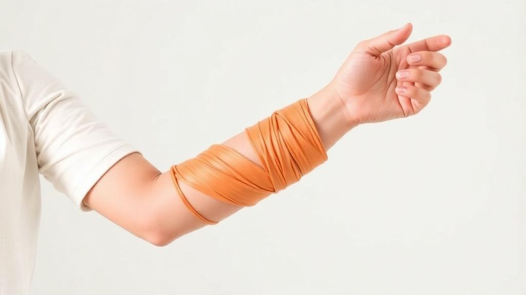
A rehabilitation technique borrowed from sports medicine — compressive wrapping of a limb with an elastic band, combined with movement — has been linked to striking functional gains in an adolescent girl with long-standing arm paralysis after a childhood stroke, according to a case report published in Frontiers in Stroke. After six months of continuous flossing intervention, the patient’s wrist extension range of motion improved from −20 degrees to 40 degrees, her clinical spasticity scores dropped across every evaluated muscle group, and she was able to fasten buttons with both hands for the first time since her stroke onset.
The case centers on a teenage girl who suffered a cerebral infarction associated with moyamoya disease — a rare condition in which arteries at the base of the brain gradually narrow — during early elementary school. She underwent cerebral revascularization surgery in the left temporal region, but postoperatively experienced a new extensive infarction involving the left cerebral hemisphere. The result was spastic paralysis predominantly affecting the distal muscles of her right upper limb: the wrist and fingers, the very structures that control grasping, releasing, and precise manipulation. Magnetic resonance imaging revealed chronic infarction extending from the left frontal lobe to part of the temporal lobe, and no alternative neurological or orthopedic condition explained her upper limb findings.
Spasticity — increased resistance to passive movement caused by disordered spinal stretch-reflex excitability after brain injury — is one of the most common and disabling sequelae of stroke. When it affects the wrist and finger flexors, it markedly limits bimanual tasks and activities of daily living. These limitations are often more pronounced in patients who experience stroke during childhood, because neurological impairment persists through critical periods of motor development. Existing treatments include botulinum toxin injections, non-invasive brain stimulation such as repetitive transcranial magnetic stimulation and transcranial direct current stimulation, and extracorporeal shock wave therapy. Although these approaches have demonstrated clinical effectiveness, their use may be constrained by limited duration of effects, the need for specialized equipment or expertise, and difficulties in maintaining long-term or home-based treatment — a particular problem for a school-age patient preparing for university entrance examinations.
Flossing intervention is a relatively novel technique that involves compressive wrapping of a limb using an elastic band, combined with passive and active movements. Originally introduced in sports and musculoskeletal rehabilitation, flossing has been reported to improve joint range of motion, reduce pain, and enhance soft tissue mobility. Proposed mechanisms include transient ischemia followed by reactive hyperemia — the surge of blood flow that follows release of compression — modulation of sensory receptors, and improved gliding of muscles, tendons, and fascia. Until now, however, clinical evidence supporting its use in patients with neurological disorders has remained limited, and there have been few reports describing its application for upper limb spastic paralysis after childhood-onset stroke.
At baseline, the patient demonstrated marked distal spasticity. The Modified Ashworth Scale, a clinical measure of resistance to passive movement, scored 1+ for her elbow flexors, 2 for her forearm pronators, and 2 for her finger flexors. Upper limb motor function assessed with the Fugl–Meyer Assessment was 43 out of 66 points, with marked impairment in distal components. Active wrist extension was limited to −20 degrees, and the thumb showed a characteristic Z deformity — adduction at the carpometacarpal joint with hyperextension at the metacarpophalangeal joint. The Motor Activity Log, which measures real-world use of the affected arm, revealed low scores for both amount of use (1.42) and quality of movement (1.71). In daily activities, the affected hand was used only as an assistive limb.
The intervention itself was technically straightforward. With the patient seated, an elastic floss band one inch in width was wrapped circumferentially around the distal forearm, extending to the elbow joint. Wrapping was initiated from the distal portion of the forearm and progressed proximally, with the band applied at approximately 1.5 times its natural length and an overlap of approximately 50 percent between layers to provide uniform compression. After wrapping, the wrapped region was manually compressed in the longitudinal direction for about 30 seconds, then twisted side-to-side for another 30 seconds. This was followed by roughly one minute of active and passive movements under compression — forearm pronation and supination, wrist flexion and extension, and finger flexion and extension. The band was then rapidly removed, followed by about ten repetitions of active movements without compression. Each set required approximately four to five minutes, and two to three sets were performed per session, for a total session duration of about eight to fifteen minutes.
Because regular outpatient attendance was difficult owing to the patient’s school schedule, supervised occupational therapy sessions were conducted approximately once every two months — roughly three supervised sessions during the six-month period. The patient’s mother was instructed in the wrapping technique and exercise procedure for implementation as a home-based program, performed at least twice daily, typically in the morning and evening, with two to three sets per session. Adherence was assessed through direct interviews and by reviewing videos of the intervention performed at home. During the six-month period, no other upper limb rehabilitation interventions were provided, the patient received no botulinum toxin injections, and no changes in medication were made. No adverse events — increased pain, skin injury, or exacerbation of neurological symptoms — were observed.
The outcomes were consistent across multiple measures. Modified Ashworth Scale scores decreased across all evaluated muscle groups: elbow flexors fell from 1+ to 0, forearm pronators from 2 to 1+, and finger flexors from 2 to 1. Fugl–Meyer upper limb scores rose from 43 to 50 out of 66. Active wrist extension improved from −20 degrees to 40 degrees — a 60-degree swing in a joint that had been frozen in flexion. Motor Activity Log scores increased for both amount of use (1.42 to 2.21) and quality of movement (1.71 to 2.64). In practical terms, the patient was able to perform bilateral hand tasks that had been impossible before the intervention, including fastening and unfastening buttons using both hands for the first time since stroke onset. She reported that her affected hand felt easier to move, and functional improvements were maintained throughout the follow-up period. At the latest follow-up, she was enjoying a fulfilling university life while actively using her affected upper limb in daily activities.
The authors propose several possible mechanisms, while emphasizing that none can be confirmed from a single case. Compressive wrapping followed by rapid removal of the band may alter local blood flow and tissue pressure, contributing to reduced resistance to passive movement and improved tissue mobility — although the authors caution that improved circulation should be regarded as a possible contributing mechanism rather than a confirmed explanation. Another potential mechanism involves modulation of sensory and proprioceptive input: compression combined with active movement under pressure may stimulate cutaneous mechanoreceptors, muscle spindles, and Golgi tendon organs, thereby altering afferent input to the central nervous system. Similar neurophysiological mechanisms have been proposed in focal vibration therapy for post-stroke spasticity, in which repetitive mechanical stimulation is thought to modulate Ia afferent input and reduce spinal stretch-reflex excitability — although the authors note that flossing and focal vibration differ in stimulus characteristics and should not be assumed to act through the same afferent pathways. Improved gliding of soft tissues, including muscles, tendons, and fascia, may also have contributed, and the increased wrist extension range of motion may reflect enhanced mechanical properties of periarticular tissues.
The authors also urge caution in interpreting the Modified Ashworth Scale reduction, because the scale assesses resistance to passive movement and does not distinguish between neural and non-neural components of increased muscle tone. The observed changes may therefore reflect not only changes in neural mechanisms but also changes in soft-tissue extensibility and mechanical resistance. No objective neurophysiological measures, such as H-reflex or F-wave recordings, were obtained, limiting mechanistic interpretation. The report’s limitations are equally clear: it describes a single case without a control condition, so a causal relationship between the intervention and the improvements cannot be established, and the potential influence of unmeasured factors cannot be excluded. The technique also carries contraindications — absolute ones include active skin infection or dermatitis, thrombosis, fractures, open wounds, heart failure, latex allergy, and anticoagulant therapy — and the authors stress that clinicians should carefully screen for these and for individual tolerance when applying compressive wrapping. In this case, the patient was not receiving medications associated with an increased risk of bleeding, and no contraindications were identified. Future studies, the authors conclude, should accumulate additional cases, evaluate the effects of flossing using controlled designs, and incorporate neurophysiological and biomechanical assessments to clarify how the technique works and which patients stand to benefit. Until then, this case stands as an intriguing signal that a simple, non-invasive, home-based wrapping technique — long a fixture of sports rehabilitation — may deserve a place in the conversation about chronic post-stroke spasticity.
Subject of Research: Case Report: Flossing intervention for chronic upper limb spasticity after childhood-onset stroke
Article Title: Case Report: Flossing intervention for chronic upper limb spasticity after childhood-onset stroke
Article References: Sasaki, S., Takahira, N., Ono, Y., Aoki, K., Kurosaki, S., Moteki, M., Fujita, R., & Kenmoku, T. (2026). Case Report: Flossing intervention for chronic upper limb spasticity after childhood-onset stroke. Frontiers in Stroke, 5, Article 1952839. https://doi.org/10.3389/fstro.2026.1952839
Image Credits: AI Generated
DOI: 10.3389/fstro.2026.1952839
Keywords: Case, Report, Flossing, intervention, chronic, upper, limb, spasticity, childhood-onset, stroke, scientific research
Cite Scienmag News
Cassandra Pierce. (October 15, 2026). Elastic Band Wrapping Shows Promise for Chronic Arm Spasticity After Childhood Stroke. Scienmag. https://scienmag.com/elastic-band-wrapping-shows-promise-for-chronic-arm-spasticity-after-childhood-stroke/
Cassandra Pierce. "Elastic Band Wrapping Shows Promise for Chronic Arm Spasticity After Childhood Stroke." Scienmag, 15 October 2026, https://scienmag.com/elastic-band-wrapping-shows-promise-for-chronic-arm-spasticity-after-childhood-stroke/. Accessed 15 October 2026.
Cassandra Pierce. "Elastic Band Wrapping Shows Promise for Chronic Arm Spasticity After Childhood Stroke." Scienmag. October 15, 2026. https://scienmag.com/elastic-band-wrapping-shows-promise-for-chronic-arm-spasticity-after-childhood-stroke/


