


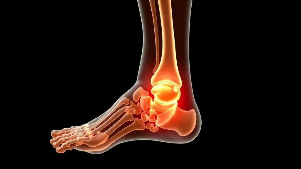
Diabetic-foot osteomyelitis, a bone infection that strikes roughly one in five people with diabetic-foot ulcers, has long been one of the most stubborn diagnostic puzzles in medicine. Now a systematic review published in the Journal of Bone and Joint Infection has taken the first comprehensive look at how machine learning is being deployed to crack the problem, and the results are both tantalizing and sobering. The review, led by Farzad Pourghazi of the Mayo Clinic together with an international team spanning the United States, Italy, Iran, and Japan, identified just ten eligible studies published between 2022 and 2026, and its findings reveal a field brimming with promise but riddled with methodological cracks that could stand between laboratory success and real-world clinical impact.
The stakes could hardly be higher. Diabetes affected an estimated 529 million adults worldwide in 2021, and between 19 and 34 percent of them will develop a foot ulcer during their lifetime. More than half of those ulcers become infected, and around 20 percent of moderate-to-severe diabetic-foot infections end in amputation. Patients with diabetic-foot ulcers face a five-year mortality rate 2.5 times higher than people with diabetes alone, and the bone infection at the center of this review drives prolonged antibiotic courses, repeated procedures, hospitalization, and limb-loss decisions. Because treatment guidelines recommend surgical resection of infected bone combined with systemic antibiotics in selected patients, an accurate diagnosis is not merely academic; it directly determines whether a limb is saved.
Why is diagnosis so hard? No single test is accurate enough on its own. Clinicians typically begin with a probe-to-bone examination, plain radiographs, and inflammatory biomarkers, escalating to magnetic resonance imaging when uncertainty persists, and reserving bone biopsy for microbiological or histopathological confirmation. Yet plain radiography has a sensitivity of only about 62 percent, and while MRI performs better, with reported sensitivity of 96.4 percent and specificity of 83.8 percent, its findings overlap with Charcot neuroarthropathy, trauma, and soft-tissue infection. The international guideline body rates the evidence for advanced imaging as only moderate, citing inconsistency and imprecision. This is precisely the kind of tangled, multi-signal problem that machine learning was built for: algorithms that can model complex relationships among clinical, laboratory, and imaging variables that no single clinician can weigh simultaneously.
The review team searched MEDLINE, Embase, Scopus, and Web of Science from January 2000 to May 2026, screening 2,343 records down to ten studies that actually developed or evaluated machine-learning models relevant to diabetic-foot osteomyelitis. Five addressed diagnosis directly: one used structured clinical and laboratory variables, two used plain radiographs, and two used MRI. The remaining studies were complementary, using osteomyelitis as a predictor within broader diabetic-foot models for wound infection or amputation outcomes, or exploring ways to make model decisions visually explainable. Critically, the authors separated these categories, refusing to let general diabetic-foot infection models masquerade as osteomyelitis diagnostic tools, a distinction that earlier reviews had blurred.
The headline numbers look spectacular at first glance. Reported discrimination, measured as the area under the receiver operating characteristic curve, ranged from 0.879 to 0.998 across direct diagnostic models, values approaching the theoretical ceiling of perfect classification. One two-center study built an explainable model from routine blood biomarkers to distinguish bone infection from soft-tissue infection alone, and it was the only direct diagnostic model subjected to independent external validation. Deep-learning models reading plain radiographs achieved image-level classification with histopathological confirmation from bone biopsy as the reference standard, while MRI-based radiomics and transfer-learning approaches differentiated osteomyelitis from Charcot neuroarthropathy and trauma with high internal discrimination.
But the review’s technical scrutiny quickly tempers the enthusiasm. Those AUC figures cannot be compared across studies because the populations, clinical endpoints, reference standards, input modalities, case mix, units of analysis, and validation designs all differed. More damning, only one of the five direct diagnostic models was ever tested on data from institutions beyond its own development cohort. Cross-validation, holdout testing, and repeated data splitting all evaluate performance on data the model has effectively seen in some form; they say nothing about whether accuracy survives a change in disease prevalence, imaging protocol, laboratory calibration, or clinical workflow. Calibration, the question of whether a model’s stated probability of infection matches reality, was reported inconsistently, and several studies omitted confidence intervals, clinically defined thresholds, or complete sensitivity and specificity figures.
The risk-of-bias assessment using the PROBAST+AI tool found most studies at high overall risk of bias, with applicability concerns in several. Imaging studies faced particular pitfalls: plain radiographs have limited sensitivity for early bone infection, so a model trained on them inherits that blind spot, and MRI studies relied on relatively small cohorts with segmented or augmented images, a known recipe for optimistic performance estimates that evaporate on new data. The reviewers also flagged the absence of any study predicting incident osteomyelitis before it develops, or modeling osteomyelitis-specific treatment outcomes such as healing, recurrence, treatment failure, amputation, or mortality. Every direct model to date has been a diagnostic classifier, not a prognostic tool.
Perhaps the most striking finding is what no one has built: a multimodal model. Clinical practice already integrates wound characteristics, probe-to-bone findings, inflammatory markers, vascular status, radiography, and MRI, yet every machine-learning study to date has fed the algorithm just one slice of that information. Clinical and laboratory variables were used without imaging; radiograph and MRI models were developed without patient-level context. The reviewers argue that future research must directly test whether combining these sources delivers incremental value, comparing clinical-only, imaging-only, and combined models on the same patient population with standardized reference standards and independent validation cohorts, rather than assuming that added complexity automatically buys better medicine.
The path forward, as the authors lay it out, demands rigor at every step: clinically available predictors gathered before the intended prediction time, careful exclusion of variables that leak the answer into the reference standard, patient-level separation during image preprocessing to prevent data leakage, external validation across institutions, calibration assessment, and decision-analytic evaluation of whether the model actually changes outcomes for patients. None of this diminishes the underlying promise. Routine blood biomarkers alone, in the one externally validated study, already distinguished bone from soft-tissue infection well enough to power a web-based clinical calculator, and deep-learning models reading ordinary X-rays hint at screening tools that could run in any clinic with a radiography suite. What the review makes clear is that the field’s next breakthrough will not come from another algorithm chasing a higher internal AUC, but from the unglamorous work of standardized endpoints, shared reference standards, and honest testing on patients the model has never met. Until then, the most sophisticated diagnostic engine for diabetic-foot osteomyelitis remains the multidisciplinary clinician, now with a growing, but still unproven, digital apprentice.
Subject of Research: Machine learning applications for diagnosing diabetic-foot osteomyelitis
Article Title: Machine learning in diabetic-foot osteomyelitis: current applications, challenges, and future directions
Article References: Pourghazi, F., Alavi, S. M. A., Petri, F., Matsuo, T., Mulett, S. A., Borgonovo, F., Gori, A., Torchia, M. E., Tande, A. J., & Berbari, E. F. (2026). Machine learning in diabetic-foot osteomyelitis: current applications, challenges, and future directions. Journal of Bone and Joint Infection, 11(5), 623-634. https://doi.org/10.5194/jbji-11-623-2026
Image Credits: AI Generated
Keywords: machine learning, diabetic-foot osteomyelitis, diabetic-foot ulcer, artificial intelligence, medical imaging, MRI, radiography, systematic review, external validation, clinical prediction models, amputation, bone infection
Cite Scienmag News
Ophelia Keating. (October 13, 2026). AI Shows Promise Against Diabetic Foot Bone Infections, But Key Gaps Remain. Scienmag. https://scienmag.com/ai-shows-promise-against-diabetic-foot-bone-infections-but-key-gaps-remain/
Ophelia Keating. "AI Shows Promise Against Diabetic Foot Bone Infections, But Key Gaps Remain." Scienmag, 13 October 2026, https://scienmag.com/ai-shows-promise-against-diabetic-foot-bone-infections-but-key-gaps-remain/. Accessed 13 October 2026.
Ophelia Keating. "AI Shows Promise Against Diabetic Foot Bone Infections, But Key Gaps Remain." Scienmag. October 13, 2026. https://scienmag.com/ai-shows-promise-against-diabetic-foot-bone-infections-but-key-gaps-remain/


