


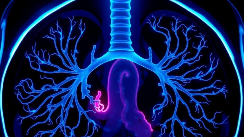
When a patient arrives at the emergency department coughing up blood, the medical team faces an urgent question: which artery in the chest is the culprit? For most cases of significant hemoptysis, the answer lies in the bronchial arteries, the small vessels that supply the lung airways and which can become enlarged and fragile in chronic inflammatory diseases such as bronchiectasis and tuberculosis. The definitive treatment is bronchial artery embolization, a procedure in which an interventional radiologist threads a catheter into the offending vessel and plugs it with particles or coils. But before that catheter ever touches the patient, doctors rely on computed tomography angiography, or CTA, to map the arterial anatomy and decide whether an enlarged artery deserves treatment. A new retrospective study from China now offers some of the most detailed numbers yet on how faithfully those CT measurements match what the angiographer actually sees on the table.
The research, published in BMC Medical Imaging by a team led by Lei Yang, Chuan Liu, Ting Li, Wen Li and corresponding author Wei He, was conducted at The First Affiliated Hospital of Chongqing Medical University. The investigators enrolled 81 patients with hemoptysis who, between June 2024 and June 2025, underwent both a bronchial artery CTA and a subsequent digital subtraction angiography, or DSA, examination before embolization. In total, 154 bronchial arteries could be analyzed in pairs, meaning that every vessel measured on the CT scan could be directly compared with the same vessel measured during the catheter study. This paired design is what gives the study its power, because it removes much of the variability that plagues comparisons between different groups of patients.
The technical question at the heart of the paper is deceptively simple: do the two imaging modalities agree? CTA is a three-dimensional, noninvasive technique in which X-ray images acquired during an intravenous contrast bolus are reconstructed into multiplanar views, allowing the radiologist to measure a vessel’s diameter at its origin and along its course. DSA, by contrast, is a two-dimensional fluoroscopic technique in which contrast is injected directly into the selected artery through a catheter. The two methods differ in physics, in timing, and in the physiological state of the vessel at the moment of imaging. A systematic difference between them could matter enormously, because clinical decisions about embolization often hinge on a single number: the widely used threshold of 2 millimeters, above which a bronchial artery is generally considered abnormally enlarged and a plausible bleeding source.
The answer from the data is reassuring at the group level. CTA-derived diameters averaged 2.34 millimeters, slightly larger than the 2.18 millimeters measured on DSA, yielding a mean vessel-level difference of just 0.16 millimeters. At the patient level, the mean difference shrank further to 0.14 millimeters. The two modalities correlated strongly, with a Spearman correlation coefficient of 0.83 and a P value below 0.001. Inter-observer reproducibility was good for both techniques, with intraclass correlation coefficients of 0.854 for CTA and 0.823 for DSA, indicating that different readers measuring the same images arrived at very similar numbers. In other words, the CT scanner is not systematically lying about the size of these arteries; it reads them, on average, a fraction of a millimeter larger than the catheter study does.
Agreement statistics, however, tell a subtler story than averages alone. The 95 percent limits of agreement, which describe the range within which most individual differences fall, spanned from minus 0.73 to plus 1.04 millimeters at the vessel level and from minus 0.55 to plus 0.83 millimeters at the patient level. That means a specific artery measured on CTA could plausibly appear up to a millimeter different from its DSA measurement. When the researchers tested the clinically meaningful 2-millimeter cutoff, using DSA as the reference standard, CTA at the same cutoff achieved a vessel-level sensitivity of 92.0 percent but a specificity of only 61.2 percent. At the patient level, the picture improved, with sensitivity of 86.7 percent and specificity of 80.4 percent. The pattern suggests that CTA tends to over-call borderline arteries: it rarely misses a truly enlarged vessel, but it sometimes flags vessels that DSA would measure just below the threshold.
For the interventionalist, this asymmetry has a practical interpretation. A CTA that shows a 2.1-millimeter bronchial artery should prompt careful interrogation during angiography rather than automatic embolization, because roughly four in ten such vessels may prove to be under 2 millimeters on direct measurement. Conversely, a CTA that shows a clearly enlarged artery of 3 or 4 millimeters can be trusted almost without reservation. The authors emphasize that CTA remains genuinely useful for preprocedural assessment, since it guides the angiographer to the relevant vessels, reveals anomalous origins such as arteries arising from the aortic arch or subclavian vessels, and shortens fluoroscopy time. What the new numbers add is a quantified sense of how much confidence to place in measurements that sit near the decision boundary.
The study also tackled a more speculative physiological question: does active bleeding at the time of imaging change the caliber of the bronchial arteries? The investigators stratified their analysis by whether the patient was experiencing hemoptysis during the CTA, during the DSA, during both, or during neither. Using a linear mixed-effects model with a random intercept per patient, a statistical approach that correctly accounts for the fact that multiple arteries from the same patient are not independent observations, they found that bleeding status at neither examination reached statistical significance. The coefficient for bleeding during CTA was minus 0.024 with a 95 percent confidence interval spanning minus 0.294 to 0.247 and a P value of 0.863, while the coefficient for bleeding during DSA was 0.057 with a confidence interval of minus 0.175 to 0.288 and a P value of 0.628. Neither comes close to conventional significance thresholds.
Yet the exploratory subgroup analysis contained an intriguing hint. When hemoptysis occurred only during the CTA, the median CTA-DSA difference was 0.00 millimeters, whereas when no bleeding occurred during either examination, the median difference rose to 0.20 millimeters at the vessel level and 0.33 millimeters at the patient level. The authors are careful to label this finding as hypothesis-generating rather than confirmatory, and they explicitly describe transient vasoconstriction as speculative. One possible mechanism they raise is that active bleeding may be accompanied by vasospasm or altered vascular tone that temporarily changes arterial caliber, potentially making the two examinations more similar if both capture the vessel in a constricted state. The subgroup in which bleeding occurred only during DSA contained just a single vessel and patient, precluding any statistical inference, and the overall between-group comparison excluding that group did not reach significance, with a Kruskal-Wallis P value of 0.154 and all Dunn-adjusted pairwise comparisons above 0.05.
The study’s limitations are those inherent to its design. It was single-center and retrospective, which means the CTA protocols and DSA techniques reflected one institution’s practice and the analysis depended on measurements already recorded in the picture archiving and communication system. The modest sample size, particularly within bleeding subgroups, limits the precision of the stratified estimates. The authors also used DeepSeek, an artificial intelligence tool, for language refinement and grammar checking, a disclosure they make transparently while taking full responsibility for the content. The work was supported by the Tibet Autonomous Region Natural Science Foundation as part of a group-style medical aid project, reflecting the collaboration between hospitals in Chongqing and the Tibetan plateau region. The ethics committee waived individual informed consent given the retrospective nature of the analysis, and the protocol was approved under the Declaration of Helsinki.
For clinicians managing hemoptysis, the takeaway is a calibrated one. CTA and DSA measure bronchial arteries with a small, predictable systematic offset of roughly 0.15 millimeters, and with strong correlation and good reproducibility, so the CT suite remains the right place to plan an embolization procedure. But the limits of agreement remind us that individual vessels near the 2-millimeter threshold can shift across the line between the two examinations, and the modest specificity of CTA at that cutoff argues for angiographic confirmation of borderline findings. As for whether the drama of active bleeding itself reshapes the arteries being measured, the data lean toward no, while leaving the door ajar for future prospective studies with larger cohorts to test the vasoconstriction hypothesis properly. In a procedure where millimeters decide treatment, knowing exactly how much the ruler can be trusted is itself a clinical advance.
Subject of Research: Comparison of bronchial artery diameter measurements between CT angiography and digital subtraction angiography in patients with hemoptysis
Article Title: Comparison of bronchial artery diameters on CT angiography versus digital subtraction angiography in hemoptysis: a retrospective study
Article References: Yang, L., Liu, C., Li, T., Li, W., & He, W. (2026). Comparison of bronchial artery diameters on CT angiography versus digital subtraction angiography in hemoptysis: a retrospective study. BMC Medical Imaging. https://doi.org/10.1186/s12880-026-02866-3
Image Credits: AI Generated
DOI: 10.1186/s12880-026-02866-3
Keywords: hemoptysis, bronchial artery, CT angiography, digital subtraction angiography, embolization, vascular measurement, interventional radiology, radiology, limits of agreement, vasoconstriction, BMC Medical Imaging, retrospective study
Cite Scienmag News
Ophelia Keating. (October 11, 2026). CT Scans Match Catheter Measurements of Bleeding Lung Arteries, Study Finds. Scienmag. https://scienmag.com/ct-scans-match-catheter-measurements-of-bleeding-lung-arteries-study-finds/
Ophelia Keating. "CT Scans Match Catheter Measurements of Bleeding Lung Arteries, Study Finds." Scienmag, 11 October 2026, https://scienmag.com/ct-scans-match-catheter-measurements-of-bleeding-lung-arteries-study-finds/. Accessed 11 October 2026.
Ophelia Keating. "CT Scans Match Catheter Measurements of Bleeding Lung Arteries, Study Finds." Scienmag. October 11, 2026. https://scienmag.com/ct-scans-match-catheter-measurements-of-bleeding-lung-arteries-study-finds/


