


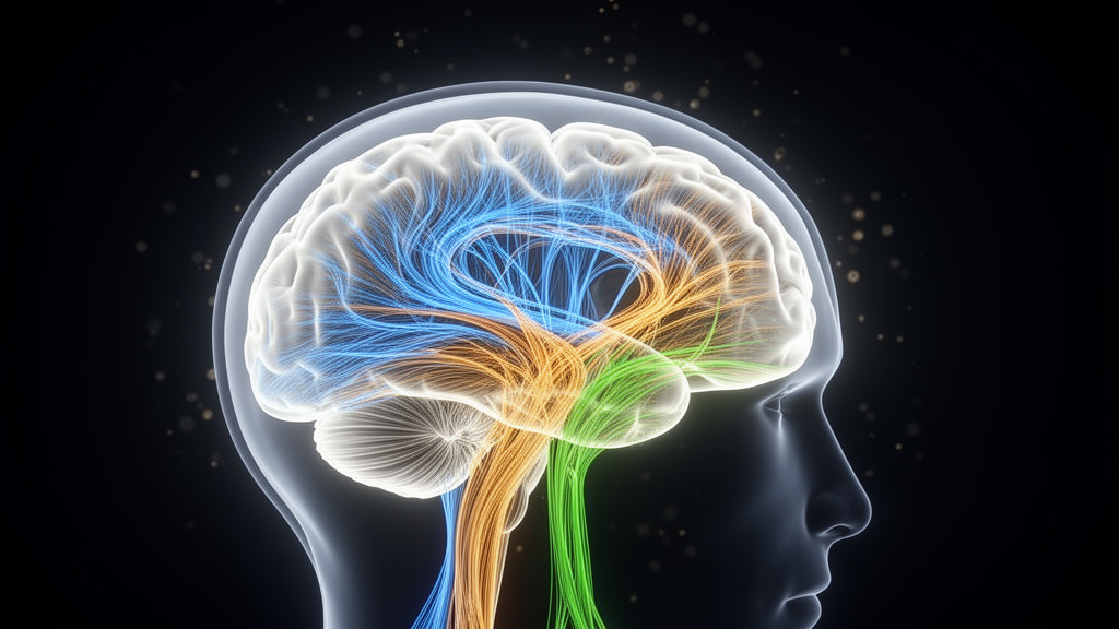
Addiction has long been treated as a single disease with many faces, but new brain imaging evidence suggests that the wiring beneath the surface tells strikingly different stories depending on the substance involved. In one of the most detailed comparisons of its kind, researchers in Germany scanned the brains of people dependent on opioids, cannabis or nicotine and found that each form of dependence leaves a distinct microstructural fingerprint in the brain’s white matter, the fiber bundles that carry signals between regions. The study, published in Translational Psychiatry, also uncovered a provocative link between the organization of a specific fiber tract and impulsive decision-making in people with cannabis dependence, a connection that held only among daily users.
The research team, led by Richard O. Nkrumah of the Central Institute of Mental Health in Mannheim and Gabriele Ende, recruited 87 individuals with substance use disorders alongside 53 non-smoking healthy controls. Participants were drawn from two clinical sites, the Central Institute of Mental Health in Mannheim and the Charité Universitätsmedizin Berlin, and all dependence diagnoses were made according to ICD-10 criteria, which correspond to severe substance use disorder under the DSM-5 framework. Crucially, the study excluded people with current comorbid substance use disorders, allowing the researchers to isolate patterns associated with each primary substance rather than muddying the picture with overlapping addictions.
What sets this investigation apart technically is its use of advanced diffusion magnetic resonance imaging, which probes the microscopic architecture of brain tissue by tracking the movement of water molecules. The team combined conventional diffusion tensor imaging, which yields measures such as fractional anisotropy and mean diffusivity, with a newer technique called neurite orientation dispersion and density imaging, or NODDI. While diffusion tensor imaging has been the workhorse of white matter research for two decades, it struggles in regions where fibers cross, which applies to as much as 90 percent of white matter. NODDI sidesteps this limitation by separately modeling neurite density, the isotropic or freely diffusing water fraction, and the orientation dispersion index, which captures how tangled or dispersed the axons within a voxel are.
The whole-brain results were unambiguous in their broad strokes. Both the opioid-dependent and cannabis-dependent groups showed widespread abnormalities characterized by reduced fractional anisotropy, elevated mean diffusivity and increased orientation dispersion across fronto-temporo-parietal and limbic pathways. Among the affected tracts were the superior longitudinal fasciculus, the arcuate fasciculus, the cingulum bundle and the middle longitudinal fasciculus, a temporo-parietal connection implicated in cognitive control and valuation. Nicotine dependence, by contrast, produced a far more circumscribed pattern, with significant alterations appearing mainly in mean diffusivity and localized to anterior and callosal white matter. The nicotine-related changes largely fell within the broader spatial footprint seen in the other two groups.
When the researchers directly overlapped the maps from the opioid and cannabis groups, they found substantial shared damage in association pathways and medial limbic tracts, but also telling differences. Opioid-dependent participants showed alterations extending further into midline and projection structures such as the corpus callosum and anterior thalamic radiation, while cannabis-related changes were more confined to fronto-parietal and temporal association fibers. These convergent and divergent signatures may reflect differences in pharmacology, chronic exposure patterns or the heavier psychiatric burden carried by the opioid and cannabis groups, who reported significantly elevated depressive symptoms and perceived stress compared with controls.
The most striking finding, however, concerned behavior. All participants completed the Kirby Delay Discounting Task, a well-validated measure of impulsivity in which people choose between smaller immediate monetary rewards and larger delayed ones. Steeper discounting, meaning a stronger preference for instant gratification, is considered a core behavioral marker of addiction. In the cannabis dependence group, higher orientation dispersion in the middle longitudinal fasciculus was significantly associated with steeper delay discounting, with a correlation of 0.41 that remained significant after controlling for age, sex, scanning site, daily alcohol consumption and nicotine dependence scores. Notably, fractional anisotropy in the same tract showed no such relationship, suggesting that the NODDI-derived measure was sensitive to microstructural features that conventional metrics missed.
The middle longitudinal fasciculus is an anatomically compelling candidate for this role. It connects the superior temporal gyrus and superior parietal lobule with dorsolateral prefrontal regions that support cognitive control and future-oriented valuation, making it a plausible conduit for integrating sensory, memory and executive inputs during decisions about delayed rewards. The researchers emphasize that orientation dispersion does not map onto any single biological process, so the association is best interpreted as altered microstructural signal within a behaviorally relevant tract rather than proof of specific fiber disorganization.
Exploratory statistical modeling added another layer of intrigue. Using a moderated mediation framework, the team found that the model explained just over half the variance in delay discounting within the cannabis group. Higher middle longitudinal fasciculus orientation dispersion predicted greater cannabis consumption, and the indirect link between tract microstructure and impulsive choice through consumption amount was significant only among daily users, not among those who used cannabis less frequently. This frequency-dependent pattern aligns with prior evidence that heavy, daily cannabis use is associated with greater disruption of fronto-parietal networks and poorer inhibitory control than occasional use. A supplementary reverse model, in which consumption severity was tested as the predictor and tract microstructure as the mediator, showed consistent associations but did not yield a statistically supported indirect effect, underscoring the exploratory nature of these analyses.
In the opioid dependence group, despite showing the most widespread white matter alterations of any group, no significant relationship emerged between middle longitudinal fasciculus measures and delay discounting. The authors caution against overinterpreting this absence, since the opioid subgroup comprised only 16 participants, all receiving opioid substitution therapy, which limits statistical power and generalizability to people with active illicit opioid use. The opioid group also carried the highest levels of perceived stress and anxiety, factors known to correlate with impulsive choice that may have obscured tract-specific associations. Opioid-related white matter abnormalities may instead involve thalamocortical and limbic-striatal circuits beyond the middle longitudinal fasciculus.
Several limitations temper the conclusions. The cross-sectional design cannot determine whether white matter abnormalities precede heavy substance use and impulsive decision-making or result from chronic exposure, a question that will require longitudinal and animal studies. Subgroup sizes were modest, particularly for nicotine and opioid dependence, and residual between-site variance in global diffusion measures persisted despite statistical harmonization. Previous, though not concurrent, polysubstance use was common, especially in the opioid group, and may have contributed unmeasured variability. Even so, the study delivers a clear message: addiction is not one white matter disease but a family of related syndromes with overlapping yet distinguishable neural signatures, and the microstructure of a single temporo-prefrontal tract may hold a measurable key to why some people with cannabis dependence struggle more than others to wait for a better reward.
Subject of Research: White matter microstructure differences across opioid, cannabis and nicotine dependence and their link to impulsive decision-making
Article Title: White matter signatures of addiction diverge across opioids, cannabis and nicotine and predict impulsive choice in cannabis dependence
Article References: Nkrumah, R. O., Wetzel, L., Böhmer, J., Eidenmueller, K., Bach, P., De Santis, S., Walter, H., Sommer, W. H., & Ende, G. (2026). White matter signatures of addiction diverge across opioids, cannabis and nicotine and predict impulsive choice in cannabis dependence. Translational Psychiatry, 16(1), Article 548. https://doi.org/10.1038/s41398-026-04491-8
Image Credits: AI Generated
DOI: 10.1038/s41398-026-04491-8
Keywords: substance use disorders, white matter, diffusion MRI, NODDI, cannabis dependence, opioid dependence, nicotine dependence, delay discounting, middle longitudinal fasciculus, impulsivity, fractional anisotropy, orientation dispersion
Cite Scienmag News
Cassandra Pierce. (October 9, 2026). Brain Wiring Tells Different Stories in Opioid, Cannabis and Nicotine Addiction, Landmark MRI Study Finds. Scienmag. https://scienmag.com/brain-wiring-tells-different-stories-in-opioid-cannabis-and-nicotine-addiction-landmark-mri-study-finds/
Cassandra Pierce. "Brain Wiring Tells Different Stories in Opioid, Cannabis and Nicotine Addiction, Landmark MRI Study Finds." Scienmag, 9 October 2026, https://scienmag.com/brain-wiring-tells-different-stories-in-opioid-cannabis-and-nicotine-addiction-landmark-mri-study-finds/. Accessed 9 October 2026.
Cassandra Pierce. "Brain Wiring Tells Different Stories in Opioid, Cannabis and Nicotine Addiction, Landmark MRI Study Finds." Scienmag. October 9, 2026. https://scienmag.com/brain-wiring-tells-different-stories-in-opioid-cannabis-and-nicotine-addiction-landmark-mri-study-finds/


