


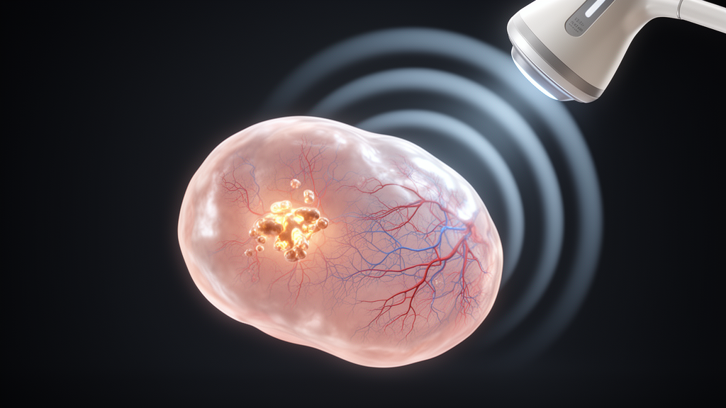
A long-standing ovarian cyst that had remained unchanged for years suddenly altered its appearance, and a new three-dimensional microvascular imaging technique helped clinicians at Peking Union Medical College Hospital uncover what conventional scans alone might have missed: a borderline ovarian tumor harboring an early-stage clear cell carcinoma. The case, published as an open-access case report in the Journal of Ovarian Research, illustrates how high-resolution visualization of tiny tumor vessels can change surgical decisions in real time, potentially preserving fertility in young women facing an ovarian cancer diagnosis.
The patient was a 35-year-old woman with a documented history of a stable ovarian cyst, a finding many women carry for years without consequence. What prompted her renewed evaluation was a recent change in the morphology of the lesion, a subtle shift in structure that raised concern despite the cyst’s otherwise unremarkable track record. Ovarian cysts are extraordinarily common, and the vast majority are benign, which is precisely why the clinical challenge is so difficult: distinguishing a harmless lesion from one that is quietly transforming into malignancy often cannot be done reliably with standard imaging alone.
To resolve the uncertainty, the clinical team deployed a multimodal imaging strategy combining ultrasound, magnetic resonance imaging, and computed tomography. All three modalities suggested malignancy, but the most decisive contribution came from three-dimensional microvascular imaging, or 3D-MVI, an advanced ultrasound technique that resolves the architecture of small blood vessels within a lesion at exceptionally high resolution. Unlike conventional color Doppler ultrasound, which detects relatively large and fast flows, microvascular imaging amplifies and maps the faint signals from slow-moving blood in capillary-scale vessels, then reconstructs them into a three-dimensional vascular map that can be rotated and interrogated from any angle.
The vascular map proved revelatory. The ultrasound examination precisely delineated the coexistence of two distinct patterns within the same ovary: features characteristic of a borderline tumor alongside vascular signatures associated with invasive malignancy. Tumor angiogenesis, the process by which a growing malignancy recruits new, architecturally chaotic blood vessels, produces recognizable patterns at the microvascular level, including irregular branching, abrupt caliber changes, and disorganized networks that differ from the more orderly vasculature of benign or low-grade lesions. Seeing both a borderline pattern and a malignant vascular pattern in a single lesion gave the clinicians an unusually specific preoperative picture of what was happening inside the cyst.
This distinction matters because of the biology of ovarian clear cell carcinoma, abbreviated OCCC. OCCC is an epithelial malignancy with a well-established association with endometriosis, and accumulating evidence indicates that it can evolve from a precursor borderline tumor. In other words, the lesion in this patient may have represented a visible snapshot of malignant transformation in progress: a borderline tumor in the process of giving rise to, or coexisting with, an invasive carcinoma. Borderline ovarian tumors themselves are not invasive cancer, and patients with pure borderline tumors generally have excellent prognoses, but the possibility of a coexisting invasive component is the scenario that clinicians most fear and most want to detect before surgery.
Armed with the microvascular findings, the multidisciplinary team made a decision that would have been difficult to justify on conventional imaging alone: they proceeded with fertility-sparing ovarian cancer staging surgery. Fertility-sparing surgery preserves the uterus and at least part of one ovary, allowing a young patient to retain the possibility of future pregnancy, but it is only appropriate when the disease is confined and the surgical staging is sufficiently rigorous to rule out spread. The preoperative confidence provided by the 3D-MVI findings, corroborated by MRI and CT, allowed the team to plan a staging procedure that balanced oncologic safety against reproductive preservation.
Histopathology after surgery confirmed the preoperative suspicion: Stage IC ovarian clear cell carcinoma coexisting with a borderline tumor. Stage IC designates disease limited to the ovary but with features such as tumor on the ovarian surface, capsular rupture, or malignant cells in ascitic fluid, placing the patient in an early but not trivially low-risk category. The concordance between the imaging prediction and the final pathology is the central lesson of the report. The ultrasound had not merely flagged generic suspicion of cancer; it had delineated the specific coexistence of borderline and malignant vascular architectures that pathology subsequently verified.
The technical achievement underlying this case deserves emphasis. Three-dimensional microvascular imaging represents a convergence of two trends in ultrasound: the development of microflow-sensitive processing that suppresses tissue motion artifacts and renders slow capillary flow visible, and volumetric acquisition that captures the vascular tree in three dimensions rather than in single planes. For ovarian lesions, which are often heterogeneous and contain solid mural components whose vascularity is the key diagnostic clue, this combination can reveal vessel patterns that two-dimensional Doppler simply cannot resolve. The authors suggest that detailed preoperative assessment using 3D-MVI offered valuable clues for detecting potential malignancy within a cyst that had appeared stable for years, and that these clues supported timely surgical decision-making, including consideration of fertility-sparing strategies.
The broader implications extend to a common clinical dilemma. Women of reproductive age with ovarian cysts face a diagnostic fork: surveillance, cystectomy, or more radical surgery, with each choice carrying trade-offs between cancer risk and reproductive or hormonal consequences. Conventional imaging cannot always distinguish a benign lesion from an early malignancy, and serum markers such as CA-125 lack specificity, particularly in early disease. A noninvasive tool that characterizes microvascular architecture could, if validated in larger studies, help stratify which stable cysts warrant closer follow-up or surgery and which can be safely observed. This report is a single case, and the authors present it as such, but it demonstrates a proof of concept: microvascular patterns can signal malignant transformation before it is otherwise detectable.
For a 35-year-old woman whose stable cyst quietly changed character, the difference between a routine cyst removal and a properly staged cancer operation may have been the resolution of a vascular map. The case, conducted under institutional ethical standards with the patient’s written informed consent and supported by China’s National Key Research and Development Program, adds to a growing body of work suggesting that the smallest vessels of a tumor carry some of the loudest diagnostic messages. As microvascular imaging matures, the hope is that fewer early ovarian cancers will hide behind the reassuring history of a stable cyst, and that more young patients will reach surgery with a diagnosis precise enough to preserve both their prognosis and their fertility.
Subject of Research: Three-dimensional microvascular ultrasound imaging for preoperative detection of malignant transformation in an ovarian borderline tumor
Article Title: 3D microvascular imaging features of an ovarian borderline tumor with coexisting clear cell carcinoma
Article References: Yang, J., Yin, J., Li, Y., Shi, X., & Su, N. (2026). 3D microvascular imaging features of an ovarian borderline tumor with coexisting clear cell carcinoma. Journal of Ovarian Research. https://doi.org/10.1186/s13048-026-02304-6
Image Credits: AI Generated
DOI: 10.1186/s13048-026-02304-6
Keywords: ovarian clear cell carcinoma, ovarian borderline tumor, microvascular imaging, 3D ultrasound, tumor angiogenesis, fertility-sparing surgery, ovarian cancer, medical imaging, endometriosis, gynecologic oncology, ultrasound, case report
Cite Scienmag News
Nathaniel Bowman. (October 9, 2026). 3D Microvascular Ultrasound Reveals Hidden Cancer in a Stable Ovarian Cyst. Scienmag. https://scienmag.com/3d-microvascular-ultrasound-reveals-hidden-cancer-in-a-stable-ovarian-cyst/
Nathaniel Bowman. "3D Microvascular Ultrasound Reveals Hidden Cancer in a Stable Ovarian Cyst." Scienmag, 9 October 2026, https://scienmag.com/3d-microvascular-ultrasound-reveals-hidden-cancer-in-a-stable-ovarian-cyst/. Accessed 9 October 2026.
Nathaniel Bowman. "3D Microvascular Ultrasound Reveals Hidden Cancer in a Stable Ovarian Cyst." Scienmag. October 9, 2026. https://scienmag.com/3d-microvascular-ultrasound-reveals-hidden-cancer-in-a-stable-ovarian-cyst/


