


Deep beneath the seafloor, the Earth keeps a written record of its own history, and scientific ocean drilling is how researchers read it. When the drilling vessel Chikyu pulls long cylinders of sediment and rock from the seabed, every meter of core is a potential archive of earthquakes, climate shifts, and ancient ocean chemistry. But before scientists can interpret these archives, they need to see inside them without breaking them apart. A team led by Mai-Linh Doan of Université Grenoble Alpes has now unveiled a deceptively simple solution to a stubborn workflow problem: a method that unwraps three-dimensional X-ray computed tomography scans of cores into flat, printable images that scientists can hold in their hands while describing the cores. The technique, developed and tested during International Ocean Discovery Program Expedition 405, is described in a technical development paper published in Scientific Drilling.
X-ray computed tomography, or XCT, has become a routine first step in core processing on board the Chikyu. The method is non-destructive, which makes it ideal for an early look at the three-dimensional structure of rocks and sediments before anyone commits to cutting, sampling, or splitting them. A medical-grade scanner, a GE Healthcare Discovery CT 750HG, is installed on the vessel. Core sections of roughly 1.5 meters are laid on a translating table and advanced through the scanner while an emitter-receiver pair rotates around them. The X-ray beam is attenuated as it passes through the sample through a combination of Compton scattering and the photoelectric effect, and the resulting attenuation coefficient is almost proportional to density. From hundreds of projections acquired at 360 degrees around the core axis, a computer reconstructs a full 3D volume that maps the density of the rock, slice by slice, from top to bottom.
The raw data, however, are unwieldy. Each core section produces DICOM files, the standard format for medical tomography, weighing several hundred megabytes apiece. Horizontal slices have a pixel size of roughly 175 micrometers, while consecutive slices are spaced 625 micrometers apart along the core axis. On board the ship, these volumes could only be explored with specialized software running on a handful of dedicated Macintosh computers located several meters from the core description tables. Scientists found themselves walking back and forth between the cores and the workstations, and checking whether a fracture or boundary was truly planar meant endlessly scrolling through parallel slices. The XCT images, in principle the most continuous and undisturbed record of the recovered material, had become a source of friction in a workflow that runs around the clock during an expedition.
Doan and colleagues propose a visual summary that strips away this friction. Their method, which they describe as unwrapping, converts the cylindrical 3D volume into a single flat 2D image, in much the same way that the Shroud of Turin is said to record a body wrapped around a cylinder. For every iso-Z slice perpendicular to the core axis, the XCT values are interpolated along a circular path centered on the core, traveling counter-clockwise to match the conventions used in borehole imaging. The circle radius is typically set to 64 pixels so that the path stays inside the core liner and samples only rock material. Recording the attenuation values along this circle for every depth produces an image in which the horizontal axis is angle around the core and the vertical axis is depth. Planar features such as fractures and lithological boundaries, which appear as complicated curves in individual slices, unfold into characteristic sinusoidal traces that trained eyes can measure directly.
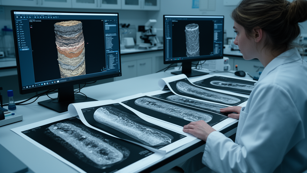
The implementation was deliberately low-tech in spirit. The team wrote Python scripts embedded in Jupyter Notebook files that read the DICOM data and generate the summary figures. On a 2020 MacBook Pro laptop, producing both the unwrapped image and the longitudinal slices of a core section took less than three minutes. The outputs were then printed on paper and delivered to two key groups: the XCT watchdog scientist, who selects whole-round core intervals for priority sampling before the cores are split, and the structural geology and sedimentology teams who conduct visual core description. Because a single color range, spanning XCT values from 0 to 3000, was applied to all cores during the expedition, the printed images also allowed scientists to track how attenuation, and therefore density, changed with depth across the entire drilled sequence.
The test bed for the method was the first window of IODP Expedition 405, carried out in late 2024 as part of the JTRACK project tracking tsunamigenic slip across the Japan Trench. The unwrapped images quickly proved their worth across the scientific party. The structure team used them to identify and measure the orientation of planar features that were difficult or impossible to distinguish from the cut surface of the core. The sedimentology team relied on them as a continuous reference for tracing fine-scale lithofacies variations, defining interval boundaries, and choosing spots for smear-slide sampling. In one striking case, a thick clast of ash resting at the bottom of the archive half, entirely hidden from the surface, would have gone unnoticed without the XCT summary. The physical properties team found that subtle variations in XCT values flagged heterogeneities throughout the core, and that distinct mineral phases such as pyrite and calcite stood out as bright, strongly absorbing regions.
Beyond the immediate operational gains, the unwrapped images opened a door to more rigorous quantitative analysis. Because they are constructed along a cylinder, they resemble the electrical and acoustic borehole images produced by downhole logging tools, and they can be loaded directly into software such as TechLog that picks sinusoidal features automatically. This allowed the team to measure the dip and relative azimuth of planar structures along the borehole faster and more precisely than manual measurements on the split working halves. Crucially, the XCT scans were collected before the cores were split, preserving the geometry of structures with minimal disturbance and yielding more reliable orientation data. Comparing unwrapped core images with borehole images also helped reorient cores where bedding could be recognized and facilitated correlating structural features between core and log, even though each dataset has limitations: borehole images are oriented and quasi-continuous but lower in resolution, while cores are high-resolution but incomplete, since recovery is rarely a full 100 percent.
The authors are careful to note that the printed XCT summaries complement rather than replace direct observation of the cores. Fine features such as slickensides, the polished fault surfaces that record slip, are invisible on X-ray images, and different lithologies can share similar densities and therefore similar XCT values, making them hard to tell apart. Visual core description at the table remains a necessity. What the unwrapping method changes is the efficiency and continuity of that description: scientists no longer need to interrupt their work to consult a distant workstation, and they can annotate a paper summary with measurements as they go, keeping the digital 3D volumes available for later, more detailed analysis.
The expedition also exposed practical hurdles. Unwrapping was not originally part of the Expedition 405 workflow, and early access to the DICOM files was not planned. Generating the images required lengthy manual copying operations so that data could be processed on an external computer disconnected from the onboard laboratory network, and this maintenance burden was high enough that usage of the summaries tended to lapse during the second window of the expedition. The authors argue that installing the Python scripts directly on a dedicated computer on board would turn XCT unwrapping into an almost real-time service running between scanning and visual core description, automatically producing depth-referenced unwrapped images and combined summaries that could also incorporate data from the Multi-Sensor Core Logger to cross-validate measurements and guide sampling.
If that integration succeeds, the humble printed image may become a standard tool of scientific ocean drilling. The method’s code and notebooks have been released openly through a Zenodo archive, lowering the barrier for other drilling platforms and even land-based core repositories to adopt the approach. For a discipline in which decisions about irreplaceable samples must often be made within hours of the core arriving on deck, a technique that turns a half-gigabyte 3D scan into a sheet of paper in under three minutes is more than a convenience. It is a way of putting the invisible interior of the Earth, quite literally, into the hands of the scientists who need to read it first.
Subject of Research: Real-time unwrapping of 3D X-ray CT scans of scientific drilling cores for visual core description
Article Title: Real-time unwrapping of 3D X-ray CT scans for visual core description
Article References: Doan, M.-L., Brunet, M., Pizer, C., Okuda, H., Chang, Y.-C., Satolli, S., Nicholson, U., Yamamoto, Y., Conin, M., Fukuchi, R., Kirkpatrick, J., Toczko, S., & the IODP Expedition 405 Scientists (2026). Real-time unwrapping of 3D X-ray CT scans for visual core description. Scientific Drilling, 35(1), 55-60. https://doi.org/10.5194/sd-35-55-2026
Image Credits: AI Generated
Keywords: X-ray computed tomography, scientific ocean drilling, IODP Expedition 405, D/V Chikyu, visual core description, core scanning, image unwrapping, structural geology, sedimentology, borehole imaging, Scientific Drilling, Japan Trench
Cite Scienmag News
Violet Maxwell. (October 9, 2026). Scientists Unwrap 3D X-ray Scans of Ocean Cores Into Paper Images in Real Time. Scienmag. https://scienmag.com/scientists-unwrap-3d-x-ray-scans-of-ocean-cores-into-paper-images-in-real-time/
Violet Maxwell. "Scientists Unwrap 3D X-ray Scans of Ocean Cores Into Paper Images in Real Time." Scienmag, 9 October 2026, https://scienmag.com/scientists-unwrap-3d-x-ray-scans-of-ocean-cores-into-paper-images-in-real-time/. Accessed 9 October 2026.
Violet Maxwell. "Scientists Unwrap 3D X-ray Scans of Ocean Cores Into Paper Images in Real Time." Scienmag. October 9, 2026. https://scienmag.com/scientists-unwrap-3d-x-ray-scans-of-ocean-cores-into-paper-images-in-real-time/


