


A 66-year-old woman arrived at her doctor’s office after coughing up roughly a cup of blood, accompanied by shortness of breath. What followed was a diagnostic journey that ended in an extraordinarily rare conclusion: transthyretin amyloidosis, a protein-misfolding disease that almost never confines itself to the airways, had been quietly depositing insoluble fibrils along her trachea and bronchi. The case, published in Respirology Case Reports, offers clinicians a vivid reminder that even well-known diseases can appear in guises so unusual they nearly escape recognition.
Amyloidosis occurs when normally soluble proteins misfold into sticky, beta-sheet-rich structures that aggregate into insoluble fibrils. These fibrils accumulate in tissues, disrupting organ function as they build up. The disease is broadly divided into systemic forms, where deposits spread throughout the body, and localized forms, where they remain confined to a single organ or tissue. Pulmonary involvement is well documented in immunoglobulin light-chain amyloidosis, the most common type affecting the lungs. But transthyretin-related amyloidosis, known as ATTR, is a different story entirely: it rarely touches the lungs at all, and when it does, its clinical features remain poorly characterized because so few cases have been described.
ATTR itself comes in two flavors. Hereditary ATTR results from pathogenic mutations in the TTR gene and runs in families. Wild-type ATTR, by contrast, is primarily an age-related phenomenon in which the normal transthyretin protein, a carrier molecule that transports thyroxine and retinol-binding protein in the blood, becomes progressively prone to misfolding over decades. In both forms, the heart, peripheral nervous system, and eyes are the organs most frequently affected. The patient in this report had no family history of amyloidosis, and genetic analysis of her peripheral blood found no pathogenic TTR variants, pointing firmly toward the wild-type, age-related variety.
When the woman was referred emergently to the hospital, her oxygen saturation had dropped and a chest radiograph revealed a new infiltrative opacity in the left mid-to-lower lung field, layered on top of a nodular opacity that had been monitored for roughly a decade. Chest computed tomography added crucial detail: bilateral bronchial wall thickening, material retained inside the left main bronchus suggestive of blood clots, infiltrative opacities concentrated in the left upper lobe, and a calcified nodule. Notably, her white blood cell count and C-reactive protein were not elevated, biochemical and coagulation parameters were normal, and tests for tuberculosis and tumor markers all came back negative, narrowing the differential diagnosis considerably.
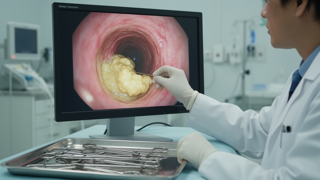
After initial stabilization with hemostatic agents, the clinical team turned to bronchoscopy, the procedure that would ultimately crack the case. The scope revealed irregular, bumpy mucosal lesions with a whitish-to-erythematous appearance extending from the trachea into both bronchi, while the peripheral airways showed diffuse wall thickening with luminal narrowing. A biopsy taken from the right upper lobe bronchial orifice, along with a bronchial aspirate, provided the tissue needed for definitive analysis. Under the microscope, pathologists found amorphous, pale eosinophilic material in the subepithelial region and around vascular walls, the classic histological signature of amyloid deposition.
Confirming the specific protein type required a sequence of staining techniques. Congo red staining, the gold-standard screen for amyloid, was positive, and the deposits resisted potassium permanganate treatment, a step that helps distinguish certain amyloid subtypes. Immunohistochemistry then delivered the decisive result: the deposits stained positive for transthyretin, establishing a diagnosis of ATTR amyloidosis. Without this layered pathological workup, the deposits could easily have been misclassified as the far more common light-chain variety, which would have sent the diagnostic workup in an entirely different direction.
Excluding systemic disease was the next painstaking task. Electrocardiography, echocardiography, and myocardial scintigraphy showed no evidence of cardiac amyloidosis, the most common and dangerous manifestation of ATTR. The patient had no sensory, motor, or autonomic nervous system symptoms, and orthopedic evaluation ruled out carpal tunnel syndrome and spinal stenosis, both frequent early signs of systemic ATTR. Ophthalmologic examination was unremarkable. To exclude multiple myeloma and other plasma cell disorders, hematologists assessed serum and urine monoclonal proteins and serum free light chains and performed a bone marrow biopsy, all of which were normal. Gastrointestinal biopsies obtained by endoscopy showed no amyloid deposition either. Collectively, these findings established that the disease was truly localized to the tracheobronchial tree, a phenotype the authors describe as exceptionally rare.
The story took one more twist when cultures of the bronchial aspirate grew Aspergillus fumigatus and Mycobacterium mucogenicum, two opportunistic organisms colonizing the damaged airway. Because both amyloid deposition and airway infection could have contributed to the hemoptysis, the team prescribed oral itraconazole for approximately one year and managed the mycobacterial infection conservatively. A CT scan performed a year later showed reduced endobronchial secretions and improved pulmonary opacities, even though the bronchial wall thickening persisted. The patient has experienced no recurrent bleeding, no progression of the airway lesions, and no emergence of systemic disease during follow-up, an encouraging outcome for a condition with no standardized treatment.
The case also carries broader lessons about how unexplained hemoptysis should be investigated. Dyspnea is the most common presenting symptom of pulmonary amyloidosis, followed by cough, weight loss, and chest pain; hemoptysis accounts for only about seven percent of presentations. Tracheobronchial amyloidosis can cause secondary infections and bleeding through mucosal fragility and bronchial stenosis driven by submucosal amyloid deposits. A prospective study of 606 patients with hemoptysis found diagnostic yields of 77.3 percent with CT, 48.7 percent with bronchoscopy, and 83.9 percent with a combined approach. In many cases, CT alone can identify the culprit, for instance when it reveals cavitary opacities strongly suggestive of tuberculosis alongside positive sputum testing. But when imaging and noninvasive tests fail to explain the bleeding, or when airway lesions are suspected, bronchoscopy can supply information nothing else can. In this patient, bronchoscopic biopsy delivered the pathological diagnosis of ATTR amyloidosis, and bronchial aspirate culture uncovered the concurrent infection that may have aggravated the bleeding, directly shaping the treatment strategy.
Treatment options for localized tracheobronchial amyloidosis remain frustratingly limited. Disease-modifying therapies for ATTR, including transthyretin stabilizers and gene-silencing agents, are approved in Japan for cardiac or neurologic involvement, but their efficacy in pulmonary disease is unknown. For airway-limited disease, interventional approaches such as bronchoscopic debulking, laser therapy, and radiotherapy have been reported, yet no standardized treatment has been established, and the evidence base rests on scattered case reports rather than controlled trials. Patients with progressive airway stenosis or recurrent hemoptysis may require such interventions, though this patient has so far been spared. The authors argue that their report demonstrates that tracheobronchial-localized ATTR can occur, however rarely, and that amyloidosis deserves a place in the differential diagnosis of hemoptysis. As wild-type ATTR becomes increasingly recognized in aging populations worldwide, clinicians evaluating unexplained airway disease may need to keep this vanishingly rare entity in mind, because the cost of missing it is measured in delayed diagnoses and unexplained bleeding that could otherwise be explained.
Subject of Research: Localized wild-type transthyretin amyloidosis of the tracheobronchial tree diagnosed after hemoptysis
Article Title: Localized Tracheobronchial Transthyretin Amyloidosis Diagnosed After Hemoptysis: A Case Report
Article References: Inoue, K., Tanaka, Y., Yanagi, M., Michimata, H., Nagahisa, Y., & Kondoh, S. (2026). Localized Tracheobronchial Transthyretin Amyloidosis Diagnosed After Hemoptysis: A Case Report. Respirology Case Reports, 14(10), Article e70781. https://doi.org/10.1002/rcr2.70781
Image Credits: AI Generated
DOI: 10.1002/rcr2.70781
Keywords: amyloidosis, transthyretin, ATTR, tracheobronchial amyloidosis, hemoptysis, bronchoscopy, Congo red staining, wild-type ATTR, pulmonary amyloidosis, airway disease, case report, respirology
Cite Scienmag News
Ophelia Keating. (October 8, 2026). Coughing Up Blood Reveals an Exceedingly Rare Form of Airway Amyloidosis. Scienmag. https://scienmag.com/coughing-up-blood-reveals-an-exceedingly-rare-form-of-airway-amyloidosis/
Ophelia Keating. "Coughing Up Blood Reveals an Exceedingly Rare Form of Airway Amyloidosis." Scienmag, 8 October 2026, https://scienmag.com/coughing-up-blood-reveals-an-exceedingly-rare-form-of-airway-amyloidosis/. Accessed 8 October 2026.
Ophelia Keating. "Coughing Up Blood Reveals an Exceedingly Rare Form of Airway Amyloidosis." Scienmag. October 8, 2026. https://scienmag.com/coughing-up-blood-reveals-an-exceedingly-rare-form-of-airway-amyloidosis/


