


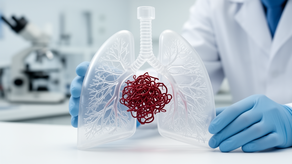
Interventional radiologists have long relied on flat, two-dimensional CT scans to navigate some of the most delicate plumbing in the human body: the tangled, abnormal blood vessels known as pulmonary arteriovenous malformations, or PAVMs. Now a multi-institution feasibility study published in the journal 3D Printing in Medicine suggests that holding a physical, patient-specific replica of those vessels in your hands before the procedure may change how these operations are planned and how patients understand what is about to happen to them. In the study, researchers at two centers specializing in hereditary hemorrhagic telangiectasia, the genetic condition that most often gives rise to PAVMs, produced three-dimensional printed models for six patients harboring eight separate malformations, and every single model was successfully fabricated and put to work in procedural planning.
The clinical problem the team set out to address is deceptively simple to describe but genuinely difficult in practice. A pulmonary arteriovenous malformation is a direct, abnormal connection between a pulmonary artery and a pulmonary vein, bypassing the capillary bed that normally sits between them. Because the capillary network usually filters small clots and bacteria out of the bloodstream, a PAVM creates a right-to-left shunt that can allow paradoxical emboli to reach the brain, causing stroke or abscess, and can produce low oxygen levels, shortness of breath, and the nosebleeds characteristic of hereditary hemorrhagic telangiectasia. The standard treatment is embolization: threading catheters through the venous system and deploying coils, plugs, or vascular occlusion devices to seal the feeding artery and shut down the malformation.
What makes embolization challenging is anatomy. PAVMs can be simple, with a single feeding artery and a single draining vein, or complex and diffuse, with multiple feeders, unusual angulation, and draining vessels that overlap confusingly on conventional imaging. The interventionalist typically works from axial CT slices, multiplanar reconstructions, and angiographic runs acquired during the procedure itself, mentally reconstructing a three-dimensional structure from two-dimensional shadows. For straightforward lesions this mental rotation is routine. For complex or recurrent malformations, particularly those in which a previous embolization attempt has failed, the cognitive load is substantial, and misjudging the angulation of a feeding vessel can cost fluoroscopy time, contrast dose, and radiation exposure for both patient and operator.
The study, led by Adam G. Fish and Anish Ghodadra at the University of Pittsburgh Medical Center together with colleagues at the University of California San Francisco and Washington University School of Medicine, took a direct approach to that problem. Using thin-slice CT angiography of each patient’s chest, the team segmented the vascular anatomy, isolating the malformation, its feeding arteries, and its draining veins from the surrounding lung parenchyma. Those segmented digital models were then sent to multimaterial printing platforms capable of rendering vessels in different colors and consistencies, so that arterial and venous structures could be distinguished at a glance and the rigid, hollow channels of the malformation could be appreciated spatially in a way no screen allows.
The technical workflow matters, because it is what makes the approach reproducible across institutions rather than a one-off demonstration. Segmentation from thin-slice CT preserves the fine detail of vessel caliber and course, which is essential when the clinical question is precisely which branch to catheterize and from which angle. Multimaterial printing, as opposed to single-resin printing, allows the model to encode additional layers of information: one material and color for the feeding artery, another for the draining vein, potentially another for surrounding structures. The fact that all six models, covering all eight malformations, were fabricated successfully is itself a meaningful result for a feasibility study, indicating that the segmentation and printing pipeline is robust enough to be deployed routinely in a clinical 3D printing laboratory rather than reserved for exceptional cases.
The payoff, according to the interventionalists who used the models, was a clearer understanding of the vascular anatomy and of the treatment approach before the patient ever reached the angiography suite. In one particularly striking case, a patient had previously undergone an attempted embolization without the benefit of a three-dimensional model, and that attempt had failed. When the procedure was planned a second time with a patient-specific printed model in hand, the embolization achieved technical success. The authors are careful about what this single case can and cannot prove; a lone anecdote cannot establish that models cause better outcomes. But as a proof of concept it captures exactly the scenario in which clinicians hope the technology will matter most: the difficult, previously treated, anatomically hostile malformation where extra spatial understanding could tip the balance between failure and success.
Across the series, embolization achieved one hundred percent technical success, meaning that in every case the operators were able to complete the intended occlusion of the malformation. The models were also used for patient education in most cases, an application that is easy to overlook but potentially transformative. PAVMs occur disproportionately in patients with hereditary hemorrhagic telangiectasia, a heritable condition that patients and families must manage for life, often involving screening of relatives, repeated imaging, and staged treatment of multiple lesions. Handing a patient a physical model of their own malformation, letting them see the feeding artery that will be plugged and the draining vein that has been stealing blood from the capillary filter, converts an abstract radiological discussion into something tangible. Informed consent becomes a conversation about a real object rather than a negotiation over gray-scale images on a monitor.
The study’s design imposes honest limits on its conclusions. It was retrospective, meaning the researchers reviewed cases that had already occurred rather than randomizing patients to model-assisted versus conventional planning. The sample, six patients and eight malformations, is the largest series reported to date for this specific application, which says as much about how young the field is as about the strength of the evidence. The reported benefits, improved operator understanding and perceived utility, are subjective assessments rather than measured endpoints. The authors themselves frame the work as demonstrating feasibility and supporting further study to define the impact of the models on procedural efficiency, clinical outcomes, and patient experience. Those future studies would need to measure hard numbers: fluoroscopy time, contrast volume, radiation dose, procedure duration, re-intervention rates, and validated patient-reported measures of comprehension and anxiety.
Even so, the findings land at a moment when three-dimensional printing is steadily migrating from engineering departments into mainstream clinical care. Patient-specific models are already used to plan complex congenital heart surgeries, to rehearse difficult neurovascular procedures, to design patient-specific implants, and to teach anatomy in medical schools. Interventional radiology has been a relative latecomer, partly because endovascular procedures are performed through small punctures with catheters rather than through open incisions, so the intuitive appeal of a physical model is less obvious than for a surgeon planning an osteotomy. Yet the cognitive challenge of navigating catheters through three-dimensional vascular space is exactly the kind of problem a tactile model can illuminate, and the Pittsburgh and UCSF results suggest that PAVM embolization is a natural fit for the technology.
The economics and logistics remain the practical frontier. Producing a patient-specific model requires image segmentation by trained personnel, access to multimaterial printers and materials, quality control, and enough lead time before a scheduled procedure, all of which carry costs that a retrospective feasibility study does not quantify. If larger prospective studies confirm that models shorten procedures, reduce radiation and contrast exposure, improve first-pass technical success in complex lesions, or measurably improve patient understanding, the case for reimbursement and routine adoption will follow. Until then, this study stands as a carefully documented demonstration that the pipeline works end to end: from thin-slice CT, through segmentation and multimaterial printing, into the hands of interventionalists and patients at two hereditary hemorrhagic telangiectasia centers, with every model successfully built and every embolization successfully completed. For a condition in which a single untreated malformation can seed a stroke, giving physicians a way to hold the problem in their hands before they fix it may prove to be more than a novelty.
Subject of Research: Patient-specific 3D-printed models for planning pulmonary arteriovenous malformation embolization
Article Title: Patient-specific 3D-printed models for pulmonary arteriovenous malformation embolization: a multi-institution feasibility study
Article References: Fish, A. G., Ghodadra, A., Bunker, M., Anwar, S., & Conrad, M. (2026). Patient-specific 3D-printed models for pulmonary arteriovenous malformation embolization: a multi-institution feasibility study. 3D Printing in Medicine. https://doi.org/10.1186/s41205-026-00346-6
Image Credits: AI Generated
DOI: 10.1186/s41205-026-00346-6
Keywords: 3D printing, pulmonary arteriovenous malformation, embolization, interventional radiology, hereditary hemorrhagic telangiectasia, CT segmentation, multimaterial printing, pre-procedural planning, patient education, vascular anatomy, HHT, medical models
Cite Scienmag News
Ophelia Keating. (October 8, 2026). 3D-Printed Lung Models Help Doctors Plug Dangerous Vessel Tangles. Scienmag. https://scienmag.com/3d-printed-lung-models-help-doctors-plug-dangerous-vessel-tangles/
Ophelia Keating. "3D-Printed Lung Models Help Doctors Plug Dangerous Vessel Tangles." Scienmag, 8 October 2026, https://scienmag.com/3d-printed-lung-models-help-doctors-plug-dangerous-vessel-tangles/. Accessed 8 October 2026.
Ophelia Keating. "3D-Printed Lung Models Help Doctors Plug Dangerous Vessel Tangles." Scienmag. October 8, 2026. https://scienmag.com/3d-printed-lung-models-help-doctors-plug-dangerous-vessel-tangles/


