


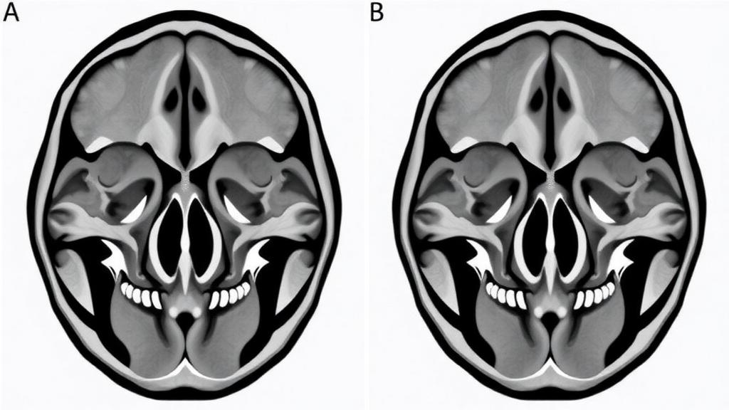
The cheekbone is one of the most recognisable features of the human face, yet the delicate seams of bone that anchor it to the rest of the skull have long resisted precise measurement. A new study published in BMC Medical Imaging has now mapped the geometry of the four zygomatic sutures in unprecedented detail, using computed tomography scans from 381 adults to establish reference values that could eventually help surgeons judge whether a shattered cheekbone has been put back together correctly. The research, led by Hilal Melis Altıntaş of Ankara Medipol University, provides the kind of large-scale, standardised anatomical baseline that the field has lacked, and its findings carry a surprise: the four sutures are far more geometrically distinct from one another than many clinicians might assume, and the symmetry between the two sides of a single person’s face is far from uniform.
The zygomatic bone, commonly known as the cheekbone, connects to four neighbouring bones through four sutures: the zygomaticomaxillary suture where it meets the upper jaw, the sphenozygomatic suture where it meets the sphenoid bone deep within the skull, the temporozygomatic suture where it meets the temporal bone, and the frontozygomatic suture where it meets the frontal bone of the forehead. Together, these junctions form the architectural framework of the zygomaticomaxillary complex, a structure that is frequently fractured in facial trauma from road accidents, sports injuries and assaults. When surgeons repair such fractures, they rely on anatomical landmarks to restore the original position of the bone, but until now there has been limited comparative data on the projected geometry of all four sutures measured within a single, consistent imaging protocol.
The research team took a retrospective cross-sectional approach, analysing cranial CT examinations that yielded 762 measurable sides across 381 adult patients. Rather than measuring the sutures directly on individual axial slices, which can distort their true course, the researchers used a technique called maximum intensity projection reformats with standardised thick slabs. This method projects the brightest bone signals along a viewing axis onto a single plane, allowing the entire winding course of each suture to be visualised and measured as a continuous line. On these projected images, the team recorded four parameters for each suture on both sides of every patient: the projected length along the suture’s full course, the end-to-end straight-line distance between its endpoints, the sinuosity, which is the ratio of the full course length to the straight-line distance and captures how winding the suture is, and the angle the suture makes with the Frankfort horizontal plane, a standard cranial reference plane that runs from the lower edge of the eye socket to the upper edge of the ear canal.
The statistical analysis was designed with care for the fact that measurements from the two sides of the same patient are not independent observations. The researchers used generalised estimating equations, a method that accounts for this within-patient correlation, and applied false discovery rate adjustment to control for the many comparisons being made simultaneously. They also calculated patient-level Spearman correlations to explore whether the dimensions of one suture predict those of another within the same individual, and intraclass correlation coefficients to quantify how closely the left and right sides of each suture matched within a single person. This combination of approaches allowed the team to distinguish genuine group-level patterns from individual variability, a distinction that proved central to their findings.
The results revealed that each of the four sutures possesses its own characteristic geometric signature. Among the defined projected measurement courses, the zygomaticomaxillary suture showed the greatest mean length and the greatest end-to-end distance, reflecting its role as the long horizontal junction between the cheekbone and the upper jaw. The orientation measurements were particularly striking: the mean angles to the Frankfort horizontal plane ranged from 15.3 degrees for the frontozygomatic suture, which runs nearly parallel to that reference plane, to 67.1 degrees for the sphenozygomatic suture, which plunges steeply into the depth of the skull. In other words, the four sutures are not variations on a theme but genuinely different structures in terms of size, winding pattern and three-dimensional orientation, each demanding its own reference values in any clinical or anatomical application.
The correlations between sutures added another layer of nuance. For dimensional parameters such as length and distance, and for sinuosity, the intersuture correlations were weak to moderate, with the largest Spearman coefficient reaching only 0.40. This suggests that a person with a long zygomaticomaxillary suture cannot be assumed to have correspondingly long other zygomatic sutures; each suture appears to develop largely on its own geometric trajectory. The angular correlations were even smaller, with the largest absolute coefficient at just 0.12, and none of the angular correlations remained statistically significant after false discovery rate adjustment. The orientation of one suture, in short, tells you essentially nothing about the orientation of its neighbours, reinforcing the picture of four independently patterned anatomical features.
Sex and side differences emerged in a pattern that clinicians will find informative. Length and end-to-end distance were greater in males for all four sutures, consistent with the overall sexual dimorphism of the craniofacial skeleton. Side differences, however, were remarkably limited at the group level: the only directional left-right difference detected was in the sphenozygomatic straight-line distance. This near-symmetry at the population level might suggest that the two sides of any individual’s face are mirror images, but the bilateral intraclass correlation coefficients told a more complicated story. These coefficients, which measure how strongly the left and right measurements correspond within the same person, ranged from 0.37 to 0.80 across the different sutures and parameters. Small average side differences, in other words, coexist with considerable individual variation in symmetry, meaning that some people have closely matched pairs of sutures while others show substantial left-right divergence even though the group averages look balanced.
This distinction between group-level symmetry and individual-level agreement has practical implications for the concept of using the uninjured side as a template for reconstructing the injured side, a common strategy in maxillofacial surgery. If bilateral agreement varies substantially from person to person, then the healthy side of a patient with a zygomaticomaxillary complex fracture may be a more or less reliable guide depending on which suture is involved and which parameter is being compared. The authors are careful to stress, however, that their data come from a single centre and a single scanner, and that the measurements were descriptive projected morphometric values obtained from a general adult population rather than from fracture patients. Before these reference values can be used in fracture reduction assessment or surgical planning, they require external validation in independent cohorts and direct evaluation in patients with actual zygomatic fractures.
The study also stands as an example of methodological transparency in modern anatomical research. The protocol was approved by the İstanbul Medipol University Non-Interventional Clinical Research Ethics Committee, and the requirement for individual informed consent was waived because of the retrospective design and the use of fully anonymised archival imaging data. The authors disclosed that a generative artificial intelligence tool was used for English-language editing, assistance with statistical coding and document formatting, but not for autonomous data analysis or interpretation, with all statistical analyses executed, reviewed and interpreted by the researchers themselves. The work received no specific external funding, and the authors declared no competing interests.
For a field in which surgical decisions can hinge on millimetre-scale judgments of bone position, the value of a large, standardised morphometric dataset is difficult to overstate. By demonstrating that the four zygomatic sutures have distinct projected profiles, that their orientations span a remarkable range from nearly horizontal to steeply inclined, and that individual bilateral symmetry is variable even when population averages appear balanced, the study gives anatomists, radiologists and surgeons a firmer quantitative foundation for understanding the midface. The next step, as the authors make clear, is to test whether these normal values can discriminate between restored and malpositioned bones in real fracture cohorts, transforming a descriptive anatomical atlas into a practical instrument for reconstructive care.
Subject of Research: Computed tomography-based morphometry of the four zygomatic sutures in adults
Article Title: Computed tomography-derived projected morphometry of four zygomatic sutures: distinct geometric profiles and bilateral agreement
Article References: Altıntaş, H. M., Köksal, A., Cingöz, G., Eberliköse, H., & Demir, B. T. (2026). Computed tomography-derived projected morphometry of four zygomatic sutures: distinct geometric profiles and bilateral agreement. BMC Medical Imaging. https://doi.org/10.1186/s12880-026-02896-x
Image Credits: AI Generated
DOI: 10.1186/s12880-026-02896-x
Keywords: computed tomography, zygomatic sutures, morphometry, craniofacial anatomy, zygomaticomaxillary complex, facial asymmetry, maxillofacial surgery, maximum intensity projection, bilateral agreement, Frankfort horizontal plane, facial trauma, BMC Medical Imaging
Cite Scienmag News
Ophelia Keating. (October 7, 2026). CT Mapping of Four Zygomatic Sutures Reveals Distinct Geometry and Uneven Left-Right Symmetry. Scienmag. https://scienmag.com/ct-mapping-of-four-zygomatic-sutures-reveals-distinct-geometry-and-uneven-left-right-symmetry/
Ophelia Keating. "CT Mapping of Four Zygomatic Sutures Reveals Distinct Geometry and Uneven Left-Right Symmetry." Scienmag, 7 October 2026, https://scienmag.com/ct-mapping-of-four-zygomatic-sutures-reveals-distinct-geometry-and-uneven-left-right-symmetry/. Accessed 7 October 2026.
Ophelia Keating. "CT Mapping of Four Zygomatic Sutures Reveals Distinct Geometry and Uneven Left-Right Symmetry." Scienmag. October 7, 2026. https://scienmag.com/ct-mapping-of-four-zygomatic-sutures-reveals-distinct-geometry-and-uneven-left-right-symmetry/


