


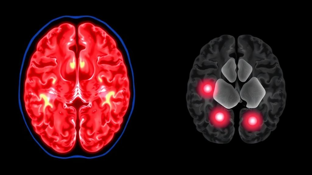
For decades, neurologists have faced an uncomfortable paradox at the bedside. A patient arrives with the classic hallmarks of a transient ischemic attack, or TIA: a sudden slurring of speech, a fleeting weakness in one arm, a momentary loss of vision that resolves within minutes. Yet when the brain is scanned with magnetic resonance imaging, the most sensitive tool available, nothing appears out of the ordinary. The attack leaves no visible trace, and the patient is sent home with a diagnosis that rests largely on clinical judgment. A new study published in the Journal of Neurology now suggests that even these seemingly invisible events leave molecular fingerprints in the blood, offering the first compelling evidence that a so-called MRI-negative TIA can still cause real, measurable damage to brain tissue.
The research, led by Francesco Berinato and colleagues at the University of Brescia and ASST Spedali Civili Hospital in Italy, focused on two proteins that have become central to the emerging field of blood-based neurology: neurofilament light chain, abbreviated NfL, and glial fibrillary acidic protein, known as GFAP. NfL is a structural component of the long, cable-like projections of neurons, and when axons are injured, fragments of this protein leak into the surrounding fluid and eventually reach the bloodstream. GFAP, by contrast, is a scaffolding protein found in astrocytes, the star-shaped support cells of the brain, and it rises when these glial cells are damaged or activated. Because each protein reports on a different cellular population, measuring both together provides a two-channel view of brain injury, capturing damage to neurons and to their supporting architecture simultaneously.
The technical challenge has always been sensitivity. Concentrations of NfL and GFAP in blood are vanishingly small, measured in picograms per milliliter, or trillionths of a gram. Conventional immunoassays simply could not detect these levels reliably in plasma. The Brescia team turned to single molecule array technology, or Simoa, an ultrasensitive digital detection platform that counts individual protein molecules by trapping them in microscopic wells studded with magnetic beads and reading out fluorescent signals one bead at a time. This approach, which has transformed research into neurodegenerative disease, made it possible to quantify subtle elevations in proteins released by microscopic, transient brain injury that no scanner can see.
The study design was deliberately conservative. The researchers enrolled 36 patients who had experienced a clinically defined TIA but whose brain MRI showed no infarct, meaning no visible stroke lesion. Each patient was matched by age and sex to two healthy controls, yielding a comparison group of 72 individuals. Blood samples were drawn within 24 hours of hospital admission, a window chosen to capture proteins while injury-related release was still at or near its peak. The team then performed group comparisons, constructed receiver operating characteristic curves to identify optimal diagnostic thresholds, and ran a series of sensitivity analyses designed to strip out confounding factors that could artificially inflate biomarker levels.
The results were striking. Patients with MRI-negative TIA had plasma NfL concentrations averaging 27.22 picograms per milliliter, more than two and a half times the control average of 10.44 picograms per milliliter, a difference that was highly statistically significant. GFAP told a similar story, averaging 301.99 picograms per milliliter in patients versus 131.65 in controls. In other words, even when the brain looked perfectly normal on imaging, the blood carried a chemical record of injury. The findings held up when the researchers systematically excluded patients with pre-existing disability, previous stroke or TIA, impaired kidney function, a heavy burden of small vessel disease on MRI, and moderate to severe cortical atrophy, all conditions known to raise NfL or GFAP independently of any acute event.
The diagnostic performance of each marker revealed an interesting asymmetry. The optimal cutoff for NfL was 16.83 picograms per milliliter, a threshold that achieved remarkable specificity of 98.6 percent, meaning that almost no healthy individual exceeded it, but sensitivity of only 52.8 percent, meaning it flagged only about half of the TIA patients. GFAP at a cutoff of 179.23 picograms per milliliter was more balanced, with sensitivity of 69.4 percent and specificity of 81.9 percent. The most powerful result emerged when the two proteins were interpreted together. A combined assessment raised sensitivity to 80.6 percent while preserving specificity at 81.9 percent, and produced the highest discriminative accuracy of any configuration tested, with an area under the curve of 0.812. The complementary biology makes sense of this: axonal injury and astroglial activation are partially independent processes, so a patient whose injury is too subtle to push one marker over threshold may still push the other.
Why does this matter clinically? TIA is fundamentally a warning sign, a brief interruption of blood flow that predicts a substantial risk of subsequent full stroke, particularly in the days and weeks that follow. Current guidelines rely on clinical scoring systems, imaging to exclude stroke mimics, and urgent vascular workup to stratify risk. But a substantial fraction of suspected TIAs remain diagnostically ambiguous, especially when imaging is negative and symptoms have fully resolved. Objective blood markers could help confirm that a transient neurological event genuinely reflected brain ischemia rather than a mimic such as migraine aura, seizure, or peripheral vestibular disorder. They could also, in principle, help quantify the tissue impact of the event, complementing what imaging cannot show.
The broader context is a field in rapid motion. Prior work has established that NfL rises acutely after overt ischemic stroke, correlates with stroke severity, and predicts functional outcome, and a recent individual patient data meta-analysis consolidated the diagnostic and prognostic value of blood NfL in ischemic stroke. GFAP has shown promise in distinguishing stroke types and mapping the timeline of tissue impact after acute cerebrovascular events. What the Brescia study adds is a crucial piece of that puzzle: evidence that the biomarker signal is not merely a byproduct of large, scan-visible infarcts, but reflects genuine microscopic injury even in the mildest end of the ischemic spectrum. This aligns with a growing recognition that TIA is not a benign non-event but a real injury whose consequences, including subtle cognitive decline after first-time TIA, may be underestimated by conventional imaging.
Important caveats remain, and the authors are careful to acknowledge them. The study was single-center and retrospective, with a modest sample of 36 patients, and the control group, while carefully matched, cannot capture the full diversity of patients who present to emergency departments with TIA-like symptoms. Specificity figures derived from healthy controls will likely look different against real-world differential diagnoses, where conditions such as renal impairment, trauma, and neurodegenerative disease can elevate the same proteins. The sensitivity analyses go a long way toward addressing these concerns, but the authors themselves state that larger studies are needed to confirm clinical utility before such tests enter routine practice.
Even so, the trajectory is clear. Blood-based biomarkers are moving from the research bench toward the stroke unit, propelled by ultrasensitive platforms like Simoa and by accumulating evidence across the cerebrovascular disease spectrum. A simple blood draw taken within a day of a suspected TIA, interpreted alongside clinical scores and imaging, could one day transform one of neurology’s most uncertain diagnoses into a measurable, quantifiable event. For patients who are told their scan is clean but their symptoms were real, the blood may soon provide the answer that the scanner cannot, confirming that even the briefest storm in the brain leaves its mark, written in the language of proteins, detectable in a few milliliters of plasma.
Subject of Research: Blood biomarkers NfL and GFAP for detecting microscopic brain injury in MRI-negative transient ischemic attack
Article Title: Blood biomarkers of brain damage in Transient Ischemic Attack: the role of plasma NfL and GFAP
Article References: Berinato, F., Morotti, A., Baronchelli, G., Tolassi, C., Girotto, I., Pilotto, A., & Padovani, A. (2026). Blood biomarkers of brain damage in Transient Ischemic Attack: the role of plasma NfL and GFAP. Journal of Neurology, 273(10), Article 580. https://doi.org/10.1007/s00415-026-14097-1
Image Credits: AI Generated
DOI: 10.1007/s00415-026-14097-1
Keywords: transient ischemic attack, TIA, neurofilament light chain, GFAP, blood biomarkers, brain injury, stroke, Simoa, MRI, neurology, cerebrovascular disease, plasma biomarkers
Cite Scienmag News
Cassandra Pierce. (October 6, 2026). Blood Test Reveals Hidden Brain Injury After Mini-Strokes With Normal Scans. Scienmag. https://scienmag.com/blood-test-reveals-hidden-brain-injury-after-mini-strokes-with-normal-scans/
Cassandra Pierce. "Blood Test Reveals Hidden Brain Injury After Mini-Strokes With Normal Scans." Scienmag, 6 October 2026, https://scienmag.com/blood-test-reveals-hidden-brain-injury-after-mini-strokes-with-normal-scans/. Accessed 6 October 2026.
Cassandra Pierce. "Blood Test Reveals Hidden Brain Injury After Mini-Strokes With Normal Scans." Scienmag. October 6, 2026. https://scienmag.com/blood-test-reveals-hidden-brain-injury-after-mini-strokes-with-normal-scans/


