


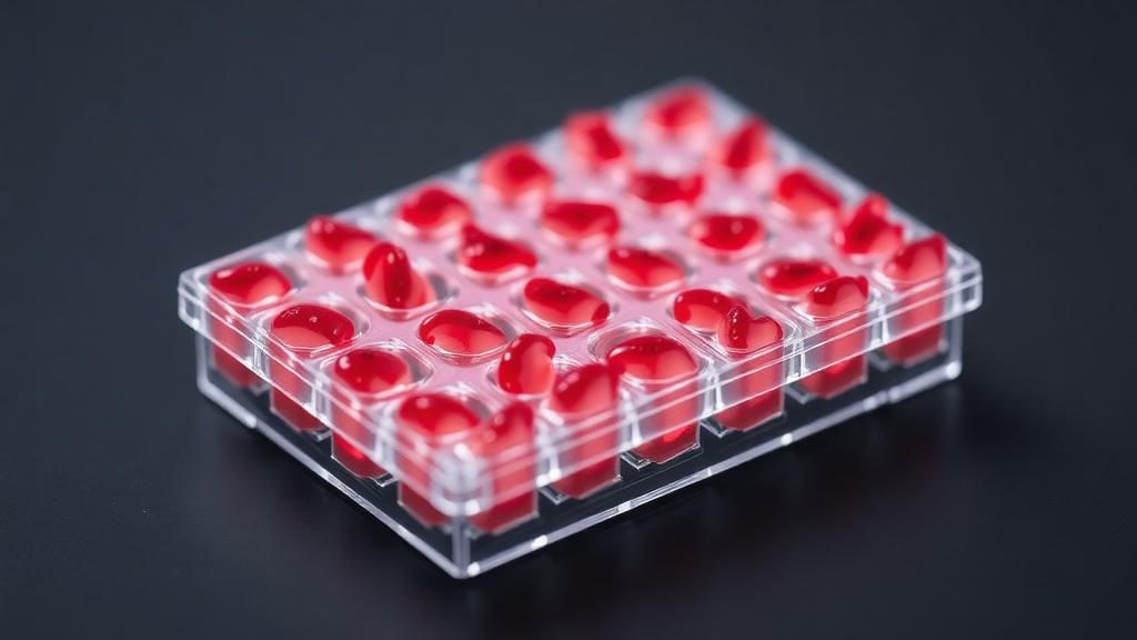
Blood vessel organoids, tiny three-dimensional clusters of human cells that recapitulate the architecture of living vasculature, have quickly become one of the most valuable tools in cardiovascular research. Now a team at the First Affiliated Hospital of Bengbu Medical University in China has reengineered how these organoids are made, moving the entire production process into a standard 96-well plate. The work, published as an open-access article in Cellular and Molecular Life Sciences, describes a scalable platform that grows one blood vessel organoid per well, allowing dozens of independent cultures to be generated, treated, and analyzed in parallel on a single plastic plate that fits in the palm of a hand.
Cardiovascular disease remains the leading cause of death worldwide, and much of that mortality traces back to vascular dysfunction, the failure of blood vessels to develop properly or to respond correctly to physiological stress. Understanding why vessels fail requires experimental models that capture the complexity of human vascular tissue, something that two-dimensional cell monolayers have never managed to do convincingly. Blood vessel organoids derived from human pluripotent stem cells address this gap by self-organizing into three-dimensional structures containing the key cellular constituents of vessel walls, providing researchers with a living miniature of human vasculature that can be studied in a dish.
The original differentiation framework that made such organoids possible was established by Wimmer and colleagues, whose protocol guided human pluripotent stem cells through a carefully choreographed sequence of developmental signals so that they matured into vascular tissue. That pioneering method, however, was built around bulk cultures in which large numbers of cell aggregates developed together in shared vessels. In such crowded conditions, neighboring cell spheres readily fuse with one another, and researchers must later perform laborious dissection to separate individual organoids. The fusion problem also means that aggregates cannot be tracked as independent experimental units, which limits the design of experiments that require many distinct conditions to be compared side by side.
The Bengbu team, led by Zhuxin Zhou, Benchi Feng, and senior authors Yong Gao and Shiyuan Chen, adapted the established differentiation framework to a U-bottom 96-well ultra-low-attachment plate. The key design choice is elegant in its simplicity: an equal number of human pluripotent stem cells is seeded into each individual well, and each well then gives rise to exactly one cell aggregate that differentiates into one blood vessel organoid. Because the U-shaped bottom of each well concentrates the cells at its center while the ultra-low-attachment surface prevents them from sticking to the plastic, the developing aggregate sits in its own private microenvironment. The one-aggregate-per-well configuration physically prevents fusion between neighboring aggregates, since each aggregate is confined to a separate well from the very start of differentiation.
This physical isolation has consequences that go far beyond convenience. Each blood vessel organoid can be cultured, treated, sampled, collected, and analyzed completely independently of its neighbors. A researcher can expose one well to a drug candidate, another to a different dose, and a third to a control vehicle, all while every organoid remains an intact, individually traceable unit. Different wells on the same plate can be assigned to distinct cell lines, treatment groups, doses, culture conditions, or time points, and the number of wells actually occupied can be adjusted freely to match the scale of any given experiment. A pilot study might use a single row of eight wells, while a full screening campaign could fill the entire plate with ninety-six independently manipulated organoids.
To demonstrate that the platform produces genuine vascular tissue, the researchers generated blood vessel organoids from two different sources of human pluripotent stem cells: the widely used H9 embryonic stem cell line and induced pluripotent stem cells, which are adult cells reprogrammed back into an embryonic-like state. They then assessed whether the resulting organoids expressed and correctly organized the molecular hallmarks of blood vessel tissue. Two complementary techniques anchored this quality control. Reverse transcription quantitative polymerase chain reaction, or RT-qPCR, measured the levels of messenger RNA for vascular marker genes, revealing which cell-type-specific genetic programs had been activated during differentiation. Immunofluorescence staining, in turn, visualized where the corresponding proteins accumulated within the organoids, confirming that vascular cells were not merely present but properly organized into vessel-like structures.
The practical advantages of the 96-well format extend to cost and labor, two factors that often determine whether an organoid method spreads through the research community. Conventional organoid protocols consume substantial quantities of expensive reagents and consumables, including type I collagen and Matrigel, the extracellular matrix mixtures used to support three-dimensional growth. Because each well in the new platform requires only the material needed for a single organoid, the total consumption of these costly components drops considerably, making the method more cost-effective for laboratories with limited budgets. The platform also eliminates much of the repetitive handling that burdens traditional workflows. The authors specifically highlight the reduction in rehanging operations, the manual transfers used to keep developing aggregates properly suspended, and the subsequent dissection work needed to separate fused organoids, saving both time and physical effort at the bench.
Flexibility is another defining feature of the system. Because plate occupancy is adjustable, the number of organoids generated can be tuned precisely to the requirements of each experiment rather than dictated by the protocol. A laboratory studying a rare patient-derived induced pluripotent stem cell line might need only a handful of organoids for a characterization study, while a pharmacology group testing a panel of vascular drugs might need hundreds across multiple plates. The platform accommodates both extremes on the same standardized hardware, which means results from different laboratories using the system should be more directly comparable than results from bespoke, hand-built organoid setups.
The researchers position the platform as a foundation for future compound-testing applications, and the logic is straightforward. Drug screening in cardiovascular research has long suffered from a shortage of human-relevant three-dimensional models that can be produced at sufficient scale and consistency. A 96-well plate is the native format of automated liquid handlers, plate readers, and high-content imaging systems, so organoids grown in this configuration slot directly into existing laboratory automation infrastructure. Multiplexed experiments, in which many compounds, doses, and genetic backgrounds are tested simultaneously, become a realistic prospect rather than an aspiration, and the ability to run distinct cell lines on a single plate opens the door to direct comparisons between healthy and disease-derived vascular tissue under identical conditions.
The work arrives at a moment when organoid technology is maturing from a specialist curiosity into a mainstream platform for disease modeling and drug development, and it addresses one of the field’s most persistent bottlenecks: throughput. By preserving the biological logic of the established differentiation framework while redesigning its physical container, the Bengbu team has created a bridge between the qualitative richness of blood vessel organoids and the quantitative demands of modern biomedical research. The study received support from the Research Fund for the Construction and Transformation of Vascular Organoid of The First Affiliated Hospital of Bengbu Medical University and from the Key Project of Natural Science Research in Universities of Anhui Province, and the article is published open access under a Creative Commons license. For laboratories worldwide that have hesitated to adopt vascular organoids because of cost, labor, or scalability concerns, the message of this platform is that the barrier to entry may now be no higher than a standard cell culture plate.
Subject of Research: A 96-well ultra-low-attachment platform for constructing human pluripotent stem cell-derived blood vessel organoids
Article Title: A 96-well platform for blood vessel organoid construction
Article References: Zhou, Z., Feng, B., Cheng, X., Zhang, X., Yu, C., Gao, Y., & Chen, S. (2026). A 96-well platform for blood vessel organoid construction. Cellular and Molecular Life Sciences. https://doi.org/10.1007/s00018-026-06388-7
Image Credits: AI Generated
DOI: 10.1007/s00018-026-06388-7
Keywords: blood vessel organoids, human pluripotent stem cells, 96-well plate, ultra-low-attachment, vascular differentiation, cardiovascular disease modeling, high-throughput screening, organoid platform, embryonic stem cells, induced pluripotent stem cells, multiplexed testing, three-dimensional culture
Cite Scienmag News
Gregory Coleman. (October 4, 2026). Mini Blood Vessels in a Miniature Plate: 96-Well Platform Streamlines Organoid Production. Scienmag. https://scienmag.com/mini-blood-vessels-in-a-miniature-plate-96-well-platform-streamlines-organoid-production/
Gregory Coleman. "Mini Blood Vessels in a Miniature Plate: 96-Well Platform Streamlines Organoid Production." Scienmag, 4 October 2026, https://scienmag.com/mini-blood-vessels-in-a-miniature-plate-96-well-platform-streamlines-organoid-production/. Accessed 4 October 2026.
Gregory Coleman. "Mini Blood Vessels in a Miniature Plate: 96-Well Platform Streamlines Organoid Production." Scienmag. October 4, 2026. https://scienmag.com/mini-blood-vessels-in-a-miniature-plate-96-well-platform-streamlines-organoid-production/


