


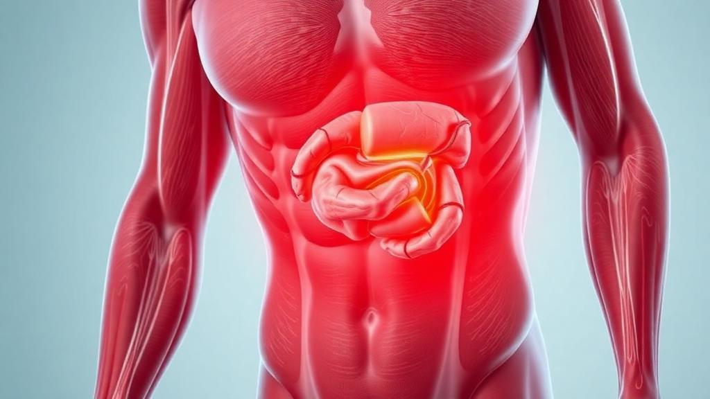
Deep inside every skeletal muscle lies a form of fat that scientists once dismissed as an incidental by-product of ageing. Today, that fat — known as intramuscular fat, or myosteatosis when it accumulates excessively — is emerging as one of the most intriguing biomarkers in medicine. A comprehensive narrative review published in the Journal of Cachexia, Sarcopenia and Muscle argues that intramuscular fat could transform how clinicians assess muscle health, predict disease outcomes and monitor treatment response, but only if the field can overcome substantial hurdles in measurement, standardisation and interpretation.
The scale of the clinical associations is striking. Excessive intramuscular fat has been linked not only to sarcopenia in older adults but also to coronary microvascular dysfunction, where it independently predicts adverse cardiovascular outcomes. It is tightly connected to insulin resistance and type 2 diabetes, with intermuscular adipose tissue shown to directly impair the insulin sensitivity of muscle tissue. In metabolic dysfunction-associated steatotic liver disease, greater intramuscular fat correlates with disease severity, visceral adiposity and even gut dysbiosis. Worsening muscle quality is associated with increased mortality in chronic kidney disease, poorer outcomes in cirrhosis, and complications after transplantation. In oncology, myosteatosis has emerged as an independent prognostic marker across multiple malignancies, predicting survival in ways that simple muscle volume measurements cannot capture.
Part of the appeal of intramuscular fat as a biomarker lies in its sensitivity to change. While ageing may cause roughly five to ten per cent muscle loss over a decade, the fat fraction within muscle can double over the same period, explaining functional decline far better than muscle mass alone. Sedentary behaviour or immobilisation quickly increases intramuscular fat content, whereas regular physical activity helps maintain lean muscle quality. Fatty infiltration in the thigh and calf muscles of older people correlates with poorer balance and increased falls risk, and in neuromuscular disorders such as muscular dystrophies, MRI fat fraction mapping has already become an invaluable outcome measure for tracking disease progression.
Yet the term myosteatosis conceals a biological complexity that the review works hard to untangle. At the microscopic level, intramyocellular lipids are stored as droplets within muscle fibres, serving as a readily mobilised energy reserve. When accumulation exceeds the storage capacity of fibres — a state known as lipotoxicity — insulin signalling is impaired and inflammatory pathways are activated. Crucially, other lipid species such as diacylglycerols and ceramides are metabolically bioactive, suggesting that lipid composition, not merely quantity, matters when interpreting the biomarker. Extramyocellular lipids occupy the space between fibres and fascicles, while intermuscular adipose tissue lies between muscle groups beneath the deep fascia. Each compartment carries different biological meaning, and the relative contribution of each to a measured fat signal depends on the imaging modality and its resolution.
That measurement challenge sits at the heart of the field. Magnetic resonance imaging offers a spectrum of approaches, from subjective visual grading systems such as the Goutallier scale, still widely used in orthopaedic surgical planning, to quantitative Dixon-based techniques that exploit the chemical shift between water and fat protons. Multi-echo Dixon sequences correct for field inhomogeneities, relaxation effects and spectral complexity, yielding the proton density fat fraction — a standardised measure validated against spectroscopy and increasingly applied in clinical trials. Magnetic resonance spectroscopy remains the gold standard for biochemical specificity, uniquely distinguishing intramyocellular from extramyocellular lipids, and phosphorus spectroscopy can probe mitochondrial energetics through high-energy phosphate metabolism. However, spectroscopy suffers from low spatial resolution, voxel placement variability and labour-intensive acquisition, confining it largely to mechanistic research.
Computed tomography offers a pragmatic alternative. It infers fat content from tissue radiographic density expressed in Hounsfield units, with lean muscle typically measuring around 50 to 60 HU and fat-infiltrated muscle appearing progressively less dense. Standardised assessment at the third lumbar vertebral level, using thresholds such as mean density below 41 HU for individuals with a body mass index under 25, has proven effective in predicting cancer survival and mortality in kidney and liver disease. Artificial intelligence, particularly convolutional neural networks, now enables fully automated segmentation of muscle and adipose tissue from routine clinical scans, reducing observer variability and opening the door to large-scale population studies. But CT carries ionising radiation, cannot separate lipid compartments, and is confounded by fluid retention and glycogen variation, which can lower muscle density independently of fat.
Ultrasound rounds out the toolkit as a portable, inexpensive, radiation-free option. Increased echogenicity indicates higher fat or fibrous tissue content, and quantitative echo intensity correlates with MRI-estimated intramuscular fat and with muscle performance. Ultrasound also captures muscle architecture — fascicle length and pennation angle — in a single bedside examination. Its weaknesses are reproducibility and specificity: measurements are sensitive to operator technique, probe pressure, transducer frequency and machine settings, and ultrasound cannot distinguish fat from fibrosis or resolve individual lipid compartments. The SARCUS consensus has defined anatomical landmarks for 39 muscles to improve acquisition consistency, and a cadaver-validated three-point grading system for sarcopenia has shown excellent reliability, but inter-device variability remains a barrier to multicentre trials.
Perhaps the most provocative finding in the review is that ‘normal’ intramuscular fat is anything but uniform. Even in healthy individuals, different muscles harbour inherently different fat content: the gluteus maximus typically contains around fifteen per cent fat in young men and twenty per cent in young women, while neighbouring hip abductor muscles hold only eight to ten per cent. Ageing amplifies these differences dramatically — quadriceps muscles in older men contain four to five times more fat than those of young men, despite only modest differences in muscle cross-sectional area. Sex differences appear partly mediated by conditioning: recreational cyclists of both sexes showed equivalent gluteal fat of roughly twelve per cent once body size and activity were accounted for. Ethnicity adds further complexity, with South Asian children carrying higher intramyocellular lipid than their peers, and African American and Hispanic adults accumulating more intramuscular fat for a given body size — patterns that may partly explain population differences in insulin resistance and that render European-derived reference values non-universal.
The athlete’s paradox crystallises why context is everything. Endurance athletes can carry high intramyocellular lipid yet remain exquisitely insulin sensitive, because their lipid droplets sit within oxidative fibres, are smaller, and are closely apposed to mitochondria, supporting rapid turnover. The same lipid levels in sedentary individuals signal insulin resistance and frailty. This U-shaped relationship — high fat in insulin-sensitive athletes, low fat in insulin-sensitive non-athletes, high fat again in insulin-resistant states — means intramuscular fat cannot be treated as a uniform risk factor. The review suggests composite indices, such as a muscle quality index integrating fat with strength or power measures, and points to emerging mass spectrometry imaging studies that map lipid species and their microregional organisation within muscle histology.
Encouragingly, intramuscular fat is modifiable. A single bout of cycling depletes intramyocellular lipid by roughly twenty-four per cent in exercised muscles. Twelve months of moderate physical activity in older adults prevented the thigh muscle fat gain seen in sedentary controls, who accumulated around eighteen per cent more fat while the active group gained only two per cent — and the exercisers avoided the strength loss their sedentary peers suffered. Meta-analyses of randomised trials show combined aerobic and resistance training reduces fat infiltration most robustly when paired with weight loss. Caloric restriction and bariatric surgery consistently reduce ectopic fat depots, though rapid weight loss risks lean mass loss unless protected by resistance training and adequate protein. Most recently, glucagon-like peptide-1 receptor agonists such as liraglutide and semaglutide have shown modest but significant reductions in intramuscular fat, though concerns persist because these drugs also reduce absolute muscle mass, raising sarcopenic obesity risks in older or frail patients. Before intramuscular fat can join bone density and liver fat as a routine clinical measure, the field needs standardised protocols, multi-ethnic longitudinal cohorts to define age-, sex- and ethnicity-specific reference ranges, and consensus on what constitutes a clinically meaningful change. If those hurdles fall, clinicians may one day stratify risk across sarcopenia, cirrhosis and cancer cachexia — and track whether an intervention genuinely improves muscle quality rather than merely mass.
Subject of Research: Intramuscular fat accumulation (myosteatosis) as a biomarker of skeletal muscle health and chronic disease
Article Title: Intramuscular Fat as a Biomarker of Muscle Health: Strengths, Limitations and Open Challenges
Article References: Spence, C., Belzunce, M. A., Di Laura, A., Henckel, J., & Hart, A. J. (2026). Intramuscular Fat as a Biomarker of Muscle Health: Strengths, Limitations and Open Challenges. Journal of Cachexia, Sarcopenia and Muscle, 17(5), Article e70389. https://doi.org/10.1002/jcsm.70389
Image Credits: AI Generated
DOI: 10.1002/jcsm.70389
Keywords: intramuscular fat, myosteatosis, sarcopenia, muscle quality, Dixon MRI, magnetic resonance spectroscopy, computed tomography, ultrasound, insulin resistance, athlete's paradox, GLP-1 receptor agonists, biomarker standardisation
Cite Scienmag News
Ophelia Keating. (October 3, 2026). Fat Hidden Inside Muscle Emerges as a Powerful New Health Biomarker. Scienmag. https://scienmag.com/fat-hidden-inside-muscle-emerges-as-a-powerful-new-health-biomarker/
Ophelia Keating. "Fat Hidden Inside Muscle Emerges as a Powerful New Health Biomarker." Scienmag, 3 October 2026, https://scienmag.com/fat-hidden-inside-muscle-emerges-as-a-powerful-new-health-biomarker/. Accessed 3 October 2026.
Ophelia Keating. "Fat Hidden Inside Muscle Emerges as a Powerful New Health Biomarker." Scienmag. October 3, 2026. https://scienmag.com/fat-hidden-inside-muscle-emerges-as-a-powerful-new-health-biomarker/


