


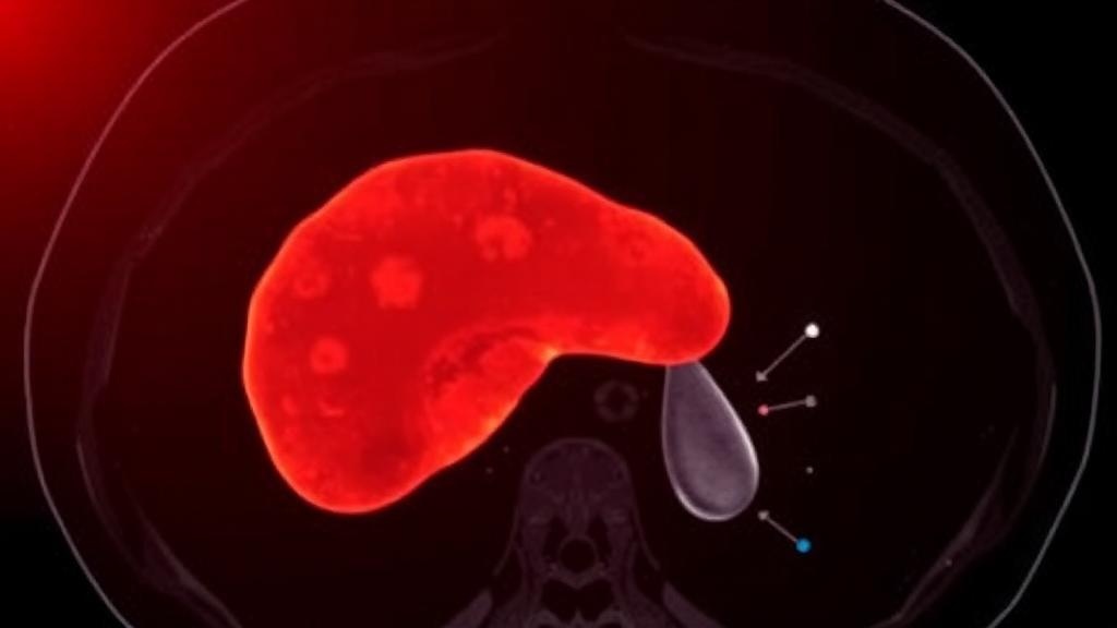
Every year, millions of abdominal computed tomography scans are performed worldwide, and a large share of them are ordered because clinicians suspect something is wrong with the liver. Reading those scans is a demanding task. Radiologists must trace the outline of an organ that blends into surrounding tissue, and then, inside it, delineate tumors that can be faint, heterogeneous, and poorly separated from healthy parenchyma. Manual contouring of the liver and its lesions is time-consuming, labor-intensive, and vulnerable to observer variability, meaning two experienced specialists can produce noticeably different outlines for the same patient. A new deep learning system described in BMC Medical Imaging aims to take over much of that burden, and its reported numbers suggest it may be one of the more capable tools yet for the job.
The system, called AFS-Net, was developed by Zheng Wang, Peng Lu, Song Liu, and Chengxin Yu of the Department of Radiology at Yichang Central People’s Hospital and the Institute of Medical Imaging at China Three Gorges University. It belongs to a family of neural network architectures known as U-shaped networks, which have become the workhorses of medical image segmentation. The name comes from their shape: an encoder that progressively compresses an image into abstract, high-level features, and a decoder that expands those features back into a full-resolution map that labels every pixel as liver, tumor, or background. The encoder captures what is in the image, while the decoder restores where it is, and the two halves exchange information through connections that span the U.
Those cross-connections, known as skip connections, are where the Chinese team concentrated their innovation. In a conventional U-shaped design, skip connections simply pass encoder features forward to the decoder, treating every piece of information as equally useful. That can be wasteful or even harmful when the features most relevant to a faint tumor boundary are drowned out by less relevant context. AFS-Net instead inserts an attention fusion module into the skip connections. The module adaptively weighs the encoder and decoder features, learning which channels and spatial locations deserve emphasis for the task at hand. In effect, the network learns to spotlight the subtle textural and contrast cues that separate tumor from liver, while suppressing features that add noise rather than signal.
The second key ingredient is a multi-scale deep supervision strategy. Training a deep segmentation network is notoriously difficult because gradients must flow backward through many layers, and early layers can receive weak or ambiguous learning signals. Deep supervision addresses this by attaching auxiliary loss functions to intermediate stages of the network, so that internal representations are also trained to be meaningful segmentations in their own right. By applying this supervision at multiple scales, AFS-Net encourages its hierarchy of features to remain faithful to the anatomy being modeled, from coarse organ-level shapes down to fine lesion boundaries. The combination of attention-based fusion and multi-scale supervision is designed to tackle precisely the challenges that have limited earlier methods: tumor heterogeneity, indistinct tumor-liver boundaries, and low-contrast lesions.
To find out whether those design choices translate into real performance, the researchers evaluated AFS-Net on the Liver Tumor Segmentation dataset, or LiTS, a widely used public benchmark of abdominal CT scans with expert-annotated liver and tumor contours. Because the data are de-identified and publicly available, the study required no direct patient recruitment or new ethics approval. The team benchmarked their model against three established competitors: 3D U-Net, ResUNet, and nnUNet, the last of which is widely regarded as a formidable, self-configuring baseline in medical segmentation. Performance was measured with three complementary metrics that capture different aspects of quality.
The first metric, the Dice similarity coefficient, measures the overlap between the automated contour and the ground truth, ranging from zero to one hundred percent. The other two metrics quantify boundary accuracy. The 95th percentile Hausdorff distance, or HD95, reports how far the predicted boundary strays from the true boundary at its worst points, using the 95th percentile to avoid being skewed by a single outlier pixel. The average symmetric surface distance, or ASSD, averages that boundary mismatch over the entire contour. Together, the three numbers tell a fuller story than overlap alone: a model can achieve a decent Dice score while still drawing clinically misleading edges, and boundary metrics expose that failure.
AFS-Net came out on top across the board. For liver segmentation, it achieved a Dice coefficient of 96.5 percent, an HD95 of 18.9 millimeters, and an ASSD of 2.1 millimeters, indicating that the organ outline was captured almost perfectly and with tight boundary agreement. Tumor segmentation is intrinsically harder, because lesions vary enormously in size, shape, and contrast, yet the network still reached a Dice coefficient of 78.2 percent, with an HD95 of 18.5 millimeters and an ASSD of 3.2 millimeters. Notably, compared with nnUNet, AFS-Net improved tumor Dice while simultaneously reducing the boundary-based error metrics, meaning the gains were not just a matter of covering more pixels but of drawing more accurate edges, which is what matters most when contours feed into treatment planning.
The authors did not rely on numbers alone. They also conducted a qualitative evaluation in which two radiologists reviewed the model’s segmentations on individual cases and judged whether the contours were acceptable. This human-in-the-loop assessment revealed a consistent pattern: contours that the radiologists accepted showed consistently better quantitative performance than those they rejected. That alignment between expert judgment and statistical metrics is important, because it suggests the metrics used in the study track what clinicians actually care about, rather than rewarding mathematical artifacts. It also provides a candid picture of where the system still falls short, since some cases were evidently rejected, a reminder that automated segmentation in the liver remains an unsolved problem at the difficult tail of the distribution.
The clinical motivation behind the work is hepatocellular carcinoma, the most common form of primary liver cancer and a disease in which accurate imaging is central to diagnosis, staging, and treatment planning. Decisions about surgery, ablation, transplantation, and locoregional therapies all depend on knowing precisely where the tumor sits and how it relates to the surrounding vasculature and organ margins. If an algorithm can produce reliable contours in seconds, it could shorten reporting times, reduce inter-observer disagreement, and serve as a consistent second reader, while still leaving the final judgment to the radiologist. The authors position AFS-Net as a support tool for computer-assisted diagnosis and treatment planning rather than a replacement for human expertise.
The study, published open access on 1 October 2026 and citable under DOI 10.1186/s12880-026-02875-2, was supported by the Beijing Medical Award Foundation, and the authors acknowledge J. Luo of Guangzhou University for guidance in code development and implementation. Like all benchmark-driven studies, it has limits that future work will need to address: the evaluation rests on a single public dataset, and performance on external clinical data, unusual tumor appearances, or post-treatment livers remains to be demonstrated. Even so, the combination of attention-guided feature fusion, multi-scale deep supervision, and strong results against a state-of-the-art baseline makes AFS-Net a noteworthy step toward segmentation tools that can genuinely ease the radiologist’s workload. As deep learning continues to move from the laboratory into the reading room, systems like this one illustrate how carefully engineered architectural choices, rather than sheer scale alone, can sharpen the machine’s eye for the subtlest lesions hidden inside a CT scan.
Subject of Research: Automated deep learning segmentation of liver and liver tumors in abdominal CT images
Article Title: A novel U-shaped AFS-Net with attention fusion module for automated liver and tumor segmentation in abdominal CT images
Article References: Wang, Z., Lu, P., Liu, S., & Yu, C. (2026). A novel U-shaped AFS-Net with attention fusion module for automated liver and tumor segmentation in abdominal CT images. BMC Medical Imaging. https://doi.org/10.1186/s12880-026-02875-2
Image Credits: AI Generated
DOI: 10.1186/s12880-026-02875-2
Keywords: liver tumor segmentation, CT imaging, deep learning, attention mechanism, AFS-Net, U-shaped network, LiTS dataset, hepatocellular carcinoma, Dice coefficient, Hausdorff distance, medical imaging, nnUNet
Cite Scienmag News
Ophelia Keating. (October 2, 2026). AI Learns to Trace Liver Tumors on CT Scans with New Attention-Powered Network. Scienmag. https://scienmag.com/ai-learns-to-trace-liver-tumors-on-ct-scans-with-new-attention-powered-network/
Ophelia Keating. "AI Learns to Trace Liver Tumors on CT Scans with New Attention-Powered Network." Scienmag, 2 October 2026, https://scienmag.com/ai-learns-to-trace-liver-tumors-on-ct-scans-with-new-attention-powered-network/. Accessed 2 October 2026.
Ophelia Keating. "AI Learns to Trace Liver Tumors on CT Scans with New Attention-Powered Network." Scienmag. October 2, 2026. https://scienmag.com/ai-learns-to-trace-liver-tumors-on-ct-scans-with-new-attention-powered-network/


