


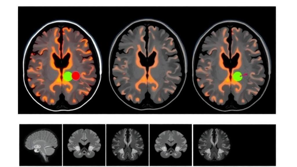
For patients whose brain metastases sit close to the skull, the difference between two medical imaging techniques may determine whether their cancer receives a full curative dose of radiation or quietly escapes it. A new dosimetric study published in BMC Cancer by Song Sun, Guanzhong Gong, Lu Zhao, and Yong Yin, based at Shandong First Medical University, Shandong Cancer Hospital, and Dalian Medical University, provides some of the most detailed quantitative evidence yet that target volumes drawn on computed tomography alone can systematically underestimate the true extent of tumors near the inner table of the skull. The finding carries immediate practical weight for radiation oncologists, because it suggests that a routine step—fusing contrast-enhanced magnetic resonance imaging with the planning CT—could prevent a meaningful fraction of lesions from being underdosed during treatment.
The research team set out to answer a deceptively simple question: when the same brain metastasis close to the skull is outlined on different imaging windows and modalities, how much do the resulting radiation dose distributions actually differ? To address it, the investigators performed a retrospective analysis of 245 lesions from 132 patients who had undergone radiotherapy. The lesions were divided among three treatment approaches: 88 treated with whole-brain radiation therapy, 67 with local radiation therapy, and 90 with a simultaneous integrated boost, a modern intensity-modulated technique that delivers a higher dose to visible tumors while treating the whole brain or surrounding tissue at a lower level simultaneously.
The methodological design is what gives the study its power. In the original clinical plans, gross tumor volumes had been delineated on contrast-enhanced brain-window CT images, a window setting optimized for brain parenchyma rather than bone. The researchers designated these volumes as GTV_Brain. They then rigidly registered contrast-enhanced MRI to the planning CT and created two additional delineations on the fused images: GTV_Skull, based on contrast-enhanced bone-window CT images that emphasize the bony structures of the skull, and GTV_MRI, based on contrast-enhanced T1-weighted MRI, which is widely regarded as the most sensitive modality for visualizing brain metastases because gadolinium contrast leaks across the disrupted blood-brain barrier of tumor tissue.
From each of the three gross tumor volumes, the team generated corresponding planning target volumes by applying a uniform three-millimeter expansion in all directions, producing PTV_Brain, PTV_Skull, and PTV_MRI. Critically, they also used Boolean operations to define MRI-only regions—the portions of the MRI-based target that extended beyond either the brain-window or bone-window CT-based targets. These regions represent tumor tissue that CT-based delineation would have missed entirely. Rather than recalculating new treatment plans, the researchers kept each patient’s original clinical plan and its dose distribution unchanged, extracting dose-volume parameters directly from the original radiotherapy dose files. This elegant approach isolates the pure effect of delineation differences on dose coverage, without confounding from plan optimization.
The primary dosimetric endpoint was D98%, the dose received by at least 98 percent of the target volume—a standard metric for confirming that nearly the entire target gets adequate coverage. The team normalized this value to the prescription dose, defining nD98% below 1.00 as a below-prescription-dose event and nD98% below 0.95 as a marked undercoverage event, a threshold beyond which a substantial portion of the tumor could harbor viable, untreated disease. Across all three treatment modalities, the pattern was strikingly consistent: PTV volumes always followed the order PTV_MRI greater than PTV_Skull greater than PTV_Brain, with the differences highly statistically significant at P less than 0.001.
The dose consequences were equally consistent and clinically sobering. D98% was highest for PTV_Brain, intermediate for PTV_Skull, and lowest for PTV_MRI. Compared with the brain-window and bone-window CT targets, the MRI-based target showed significantly lower D98% values, with reductions of 0.68 Gy and 0.58 Gy respectively in the whole-brain radiation therapy group, 1.96 Gy and 1.90 Gy in the local radiation therapy group, and 3.16 Gy and 2.82 Gy in the simultaneous integrated boost group. In other words, the more conformal and dose-escalated the technique, the greater the penalty for missing tumor tissue during delineation. A technique that concentrates radiation tightly around a drawn target is unforgiving of any tissue left outside that target.
The incidence figures translate these Gy-level differences into patient-level risk. Below-prescription-dose events for the MRI-based target occurred in 22.73 percent of lesions treated with whole-brain radiation therapy, 35.82 percent with local radiation therapy, and a striking 62.22 percent with simultaneous integrated boost. Marked undercoverage—nD98% falling below 0.95—affected 6.82 percent, 17.91 percent, and 31.11 percent of lesions in those same groups. Most alarming of all, within the MRI-only regions themselves, marked undercoverage reached 41.11 percent and 42.22 percent in the simultaneous integrated boost group, depending on which CT-based reference target was used. Nearly half of the tumor tissue visible only on MRI sat in low-dose zones that the original plans never intended to treat at full prescription.
Why does this matter biologically? A region of gross tumor that receives less than the prescription dose—and especially less than 95 percent of it—represents a geographic miss, a pocket of viable cancer cells that survives the treatment course. For brain metastases, such residual disease can lead to local recurrence, neurological deterioration, and the need for repeat radiation or surgery, all of which carry substantial morbidity. The skull-adjacent location compounds the problem: lesions hugging the inner table of the skull are precisely where contrast enhancement on CT is hardest to distinguish from adjacent bone and dural structures, and where the steep dose gradients of modern conformal techniques leave the least margin for error. The study’s data show that bone-window CT, while better than brain-window CT at capturing tumor extent near bone, still falls short of MRI.
The technical explanation lies in the physics of the two modalities. CT excels at depicting electron density, which is why it remains the backbone of dose calculation, but its soft-tissue contrast is limited, and beam-hardening artifacts near bone can obscure subtle enhancement. T1-weighted MRI after gadolinium injection, by contrast, highlights areas where the blood-brain barrier has broken down, revealing tumor margins that are invisible or ambiguous on CT. When the researchers fused the two datasets and compared delineations, the MRI-referenced targets were consistently larger, and the extra volume—the MRI-only regions—tended to lie in areas that the original plans had deliberately spared, such as regions near the scalp, skull, and normal brain that planners had constrained to keep doses low.
The authors’ conclusion is direct and actionable: for brain metastases close to the skull treated with local radiation therapy or simultaneous integrated boost, routine fusion of contrast-enhanced MRI with the planning CT is recommended for gross tumor volume delineation to reduce the potential risk of target geographic miss. The study, approved by the ethics committee of the Affiliated Cancer Hospital of Shandong First Medical University and conducted with written informed consent from all patients, was supported by research programs in Shandong Province and the Xinjiang Uygur Autonomous Region. As intensity-modulated radiotherapy, volumetric modulated arc therapy, and stereotactic techniques continue to push dose gradients ever steeper, this work serves as a quantitative reminder that the sharpest treatment plan is only as good as the contour it is built on—and that for tumors pressed against the skull, the contour drawn without MRI may simply be the wrong one.
Subject of Research: Dosimetric comparison of CT-based and MRI-based target delineation in radiotherapy for brain metastases close to the skull
Article Title: Dosimetric analysis of CT/MRI-based target delineation differences in radiotherapy for brain metastases close to the skull
Article References: Sun, S., Gong, G., Zhao, L., & Yin, Y. (2026). Dosimetric analysis of CT/MRI-based target delineation differences in radiotherapy for brain metastases close to the skull. BMC Cancer. https://doi.org/10.1186/s12885-026-17067-y
Image Credits: AI Generated
DOI: 10.1186/s12885-026-17067-y
Keywords: brain metastases, radiotherapy, target delineation, MRI, computed tomography, dosimetry, D98%, gross tumor volume, planning target volume, simultaneous integrated boost, geographic miss, radiation oncology
Cite Scienmag News
Nathaniel Bowman. (October 2, 2026). MRI Fusion Reveals Hidden Radiation Targets in Skull-Adjacent Brain Metastases. Scienmag. https://scienmag.com/mri-fusion-reveals-hidden-radiation-targets-in-skull-adjacent-brain-metastases/
Nathaniel Bowman. "MRI Fusion Reveals Hidden Radiation Targets in Skull-Adjacent Brain Metastases." Scienmag, 2 October 2026, https://scienmag.com/mri-fusion-reveals-hidden-radiation-targets-in-skull-adjacent-brain-metastases/. Accessed 2 October 2026.
Nathaniel Bowman. "MRI Fusion Reveals Hidden Radiation Targets in Skull-Adjacent Brain Metastases." Scienmag. October 2, 2026. https://scienmag.com/mri-fusion-reveals-hidden-radiation-targets-in-skull-adjacent-brain-metastases/


