


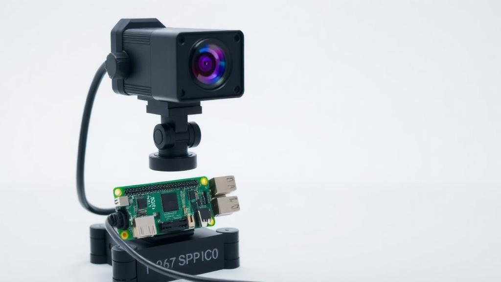
A team of biomedical engineers has built a complete scaffold-inspection system out of a Raspberry Pi camera, a ring of LEDs and some 3D-printed plastic, and shown that it measures pores in tissue-engineering scaffolds far more consistently than experienced human operators. The platform, described in Medical & Biological Engineering & Computing, costs roughly €170 in hardware and reduces the operator-to-operator variability that has long plagued manual pore measurement in tissue engineering laboratories. For a field where the geometry of a scaffold can decide whether bone regrows or fails to form, an affordable and repeatable quality-control tool could change how laboratories validate the constructs they print every day.
The problem the researchers set out to solve is deceptively simple to state. In tissue engineering, scaffolds are the porous structures that support cell attachment and guide new tissue formation, and in craniofacial applications, where surgeons aim to reconstruct complex bones of the skull and face, the pore size, shape and spatial distribution of a scaffold are decisive for regenerative success. Additive manufacturing has made it possible to print scaffolds with predefined porosity, but what comes out of the printer does not always match what went into the design file. Bioinks, the cell-laden hydrogel mixtures used in bioprinting, can shear, spread or rearrange during extrusion, degrading shape fidelity. Verifying the printed result has traditionally meant laborious manual image analysis in software such as ImageJ or CellProfiler, an approach that is slow and, crucially, vulnerable to user bias.
The new platform couples custom 3D-printed hardware with a dedicated image-processing pipeline written in MATLAB. The imaging rig consists of a 12.3-megapixel Raspberry Pi High Quality Camera built around the Sony IMX477 sensor, fitted with a 6 mm wide-angle CS-mount lens and mounted on an adjustable 3D-printed monopod. A NeoPixel 12-LED RGB ring, held in a flexible thermoplastic polyurethane adapter, bathes the sample in homogeneous white light against a high-contrast black-and-white background, sharpening the contours of scaffold edges and suppressing the shadows that would otherwise corrupt segmentation. A Raspberry Pi 3 Model B+ and an Arduino Uno handle communication between the camera, the lighting and the host computer, and the structural parts of the mount were printed in polylactic acid, the workhorse polymer of desktop 3D printing.
The software side is where the engineering becomes genuinely distinctive. The pipeline takes a single zenithal image of the scaffold and processes it through a fixed sequence of operations: rotation, correction of the barrel distortion introduced by the wide-angle lens using a radial distortion model with an empirically determined coefficient of −0.15, grayscale conversion, interactive cropping, and automated segmentation by Otsu’s global thresholding, which chooses the black-and-white cutoff that maximises inter-class variance in the image histogram. Connected-component labelling with 8-connectivity then identifies each pore, and blob analysis filters out objects that are too small or too large to be genuine pores. The system outputs the number, area, perimeter and compactness of every pore in the uppermost printed layer, exporting the results to a spreadsheet alongside an annotated overlay image.
One of the most elegant technical contributions is the compactness metric. Rather than using the classical isoperimetric quotient, which anchors a circle at unity, the authors square-normalise the measure so that a perfect square pore scores exactly one, matching the orthogonal geometry that most printed scaffolds are designed to have. A penalty function then converts deviations in either direction, pores that round off or become convoluted, into a percentage quality score between 0 and 100 percent, with a perfect square yielding 100 percent and a perfectly circular pore scoring 72.7 percent. The authors note that their metric is the reciprocal of the printability index commonly used to assess bioink shape fidelity, which allows their values to be compared directly with the existing bioprinting literature. A colour-indexed graph maps each pore’s score onto a gradient, giving researchers an at-a-glance map of where a print went wrong.
The validation strategy was deliberately staged across four assays. First, a printed reference grid with known 0.5 cm square cells established the system’s trueness and repeatability. The platform proved remarkably repeatable, with a coefficient of variation of just 1.33 percent, but it systematically underestimated pore area by 11.6 percent, measuring a mean of 0.221 square centimetres against a nominal 0.250. The authors trace this bias to the manual pixel-per-centimetre calibration and to the segmentation threshold placing the detected edge slightly inside the true pore boundary. Because the error is systematic and reproducible, it can be removed by calibrating against a reference of certified area, a straightforward fix that turns a flaw into a documented, correctable offset.
The second assay delivered the headline comparison. On a 3D-printed polylactic acid scaffold containing three classes of pores, five repeated acquisitions by the platform agreed to within a coefficient of variation of 0.01 to 2.48 percent. The same specimen was then measured manually by three experienced tissue engineering researchers, and the results were striking: inter-user coefficients of variation ranged from 11.5 to 22.8 percent, and intra-user variability from 0 to 12.7 percent. Counterintuitively, the manual measurements diverged most on the larger pores, the opposite of the pixel-resolution sensitivity shown by the automated system on small features. The authors argue this exposes a systemic problem in collaborative laboratories, where measurement quality depends on individual attention and experience, and where dispersion between operators can exceed an order of magnitude.
Feasibility was then demonstrated on materials far harder to image than rigid plastic. Two self-setting silica–gelatin hybrid bioinks, differing only in their gelatin-to-sol volume ratio, were printed into 16-pore scaffolds on a Cellink BioX bioprinter; because the inks are transparent, they were dyed with methylene blue to create contrast. The software successfully segmented the complex, non-linear pore boundaries of these hydrated hydrogel constructs, where manual measurement is most error-prone, and revealed a printing resolution error of 0.0051 square centimetres relative to the design ground truth. A final assay on a brittle silica-based scaffold showed the compactness output flagging shape deviations that the printer’s settings were supposed to prevent. The authors are careful to note that these last assays were feasibility demonstrations without independent reference measurements, not full accuracy validations.
The platform does not attempt to replace micro-computed tomography, the gold standard for resolving a scaffold’s internal three-dimensional architecture, pore interconnectivity and through-thickness geometry. Micro-CT remains expensive, slow, with acquisition and reconstruction reaching 19.5 hours and 166 gigabytes per specimen at the finest pixel sizes, and it introduces dehydration and staining artefacts in hydrated hydrogels. The optical platform instead quantifies the two-dimensional projected macrotopography of the top printed layer, positioning itself as a rapid, non-destructive screening complement to volumetric imaging. Its limitations are candidly acknowledged: the workflow is semi-automated, with focus, aperture, monopod height, illumination and crop region set by the operator, the distortion coefficient must be redetermined for any different optical configuration, and low-contrast specimens may require staining.
What makes the work resonate beyond its immediate niche is its accessibility. The entire bill of materials comes to approximately €170 excluding VAT, the source code and 3D-printable STL files are openly available under an MIT licence, and the pipeline relies only on standard image-processing primitives, meaning it can be reproduced without a commercial MATLAB licence using GNU Octave or Python with OpenCV. By demonstrating that repeatability can be made independent of operator experience, the team offers laboratories a practical route to objective quality control in scaffold fabrication. The authors emphasise that no craniofacial-specific or clinical specimen was evaluated in this study, and that establishing utility in that demanding setting will require dedicated validation. But as patient-specific scaffolds for skull and facial bone reconstruction move closer to the clinic, the ability to verify, cheaply and reproducibly, that what was printed matches what was designed is exactly the kind of unglamorous infrastructure that turns promising biofabrication into reliable medicine.
Subject of Research: A low-cost semi-automated imaging platform for quantitative characterisation of 3D-printed scaffold surface macrotopography in tissue engineering
Article Title: A low-cost imaging platform for quantitative characterisation of scaffold surface macrotopography with potential application in craniofacial tissue engineering
Article References: Marimon, X., Saman-Sakkal, E., Rodriguez, R., Portela, A., Cerrolaza, M., Mateos, M. A., & Pérez, R. (2026). A low-cost imaging platform for quantitative characterisation of scaffold surface macrotopography with potential application in craniofacial tissue engineering. Medical & Biological Engineering & Computing. https://doi.org/10.1007/s11517-026-03661-6
Image Credits: AI Generated
DOI: 10.1007/s11517-026-03661-6
Keywords: tissue engineering, scaffolds, 3D printing, bioprinting, image processing, pore analysis, craniofacial regeneration, Raspberry Pi, shape fidelity, quality control, bioinks, computer vision
Cite Scienmag News
Denise Maddox. (October 1, 2026). €170 Raspberry Pi Camera System Tames Human Error in 3D-Printed Scaffold Quality Control. Scienmag. https://scienmag.com/e170-raspberry-pi-camera-system-tames-human-error-in-3d-printed-scaffold-quality-control/
Denise Maddox. "€170 Raspberry Pi Camera System Tames Human Error in 3D-Printed Scaffold Quality Control." Scienmag, 1 October 2026, https://scienmag.com/e170-raspberry-pi-camera-system-tames-human-error-in-3d-printed-scaffold-quality-control/. Accessed 1 October 2026.
Denise Maddox. "€170 Raspberry Pi Camera System Tames Human Error in 3D-Printed Scaffold Quality Control." Scienmag. October 1, 2026. https://scienmag.com/e170-raspberry-pi-camera-system-tames-human-error-in-3d-printed-scaffold-quality-control/


