


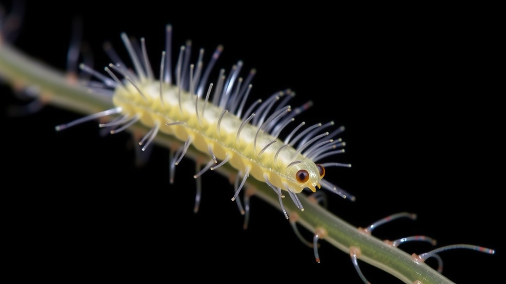
A tiny parasitic flatworm larva has rewritten one of the textbook rules of its own lineage, and the way it did so is turning heads among evolutionary biologists. Digenean trematodes, a hugely successful group of parasitic flatworms, begin life as miracidia, ciliated swimming larvae whose beating surface hairs help them hunt down a snail host. In many species these larvae are graceful, actively propelled swimmers covered in bands of cilia. But a new study of the hemiuroid trematode Bunocotyle progenetica reveals a larva that has abandoned ciliation almost entirely, replacing its ciliated surface with an armor of spines, and in doing so has co-opted one of the most recognizable components of the cilium itself as the structural backbone of its new exterior.
The research, carried out by Peter A. Smirnov, Alexandra N. Ivanova and Anna Gonchar and published in Frontiers in Zoology, combines serial transmission electron microscopy with experimental infection of the snail host to reconstruct, in remarkable detail, what happens when a larva miniaturizes and changes its infection strategy. The findings show that the spines covering the miracidium of B. progenetica are not simply modified cilia. Instead, each spine is supported internally by an elongated intracellular structure that closely resembles the striated rootlet of a cilium, the anchoring apparatus that normally tethers cilia into the cell body. In effect, the parasite appears to have dismantled the cilium and repurposed its rootlet as a scaffold for a completely different surface structure.
This kind of repurposing, known to evolutionary biologists as exaptation, is one of the most intriguing mechanisms by which novel traits arise. A structure that evolved for one function, in this case anchoring and supporting motile cilia, is recruited for an entirely new role, here providing mechanical support for spines on the larval body surface. Because larval ciliation is considered one of the defining features of the Neodermata, the larger clade that includes trematodes, tapeworms and roundworms, its complete loss in B. progenetica is a striking departure. The new study suggests that such a transformation is not only possible but can be traced at the ultrastructural level to a specific recycling of ciliary components.
The context for this transformation lies in how the larva reaches its host. In most digeneans, miracidia are free-swimming and must actively locate and penetrate a mollusc. That lifestyle demands cilia, sensory equipment and a muscular, coordinated body. But in several digenean lineages, including the Hemiurata group to which B. progenetica belongs, the miracidium has been miniaturized and has switched to a passive strategy: instead of swimming to find a snail, it simply waits to be swallowed. Once inside the digestive tract of the mollusc, it needs no cilia for locomotion, but it may well benefit from a surface that can withstand the mechanical and chemical rigors of the gut environment. Spines, the authors argue, fit that bill.
Using serial transmission electron microscopy, the team reconstructed the body wall of the miracidium in three dimensions and found it covered by three spiny epithelial plates. This is itself unusual; the neodermis, the syncytial outer covering characteristic of neodermatan parasites, is typically organized into distinct cytoplasmic regions, and its precise architecture varies across lineages. In B. progenetica, the plates carry spines across the entire body surface, an extreme condition even among spined hemiuroid miracidia, many of which bear spines only on restricted regions of the body. The internal support of each spine by a striated-rootlet-like structure suggests a developmental pathway in which the machinery that once built cilia has been redirected toward building spines.
Miniaturization has affected far more than the surface. Compared with the miracidia of non-miniaturized digeneans, which can be relatively large and anatomically elaborate, the miracidium of B. progenetica shows marked reduction across all of its organ systems. Nervous elements, musculature, excretory structures and other components are all simplified. This pattern is consistent with a broader trend in which passive infection relieves the larva of the need for the complex equipment of an active swimmer. What remains is a streamlined infective stage whose principal external features, the spines, reflect its new route into the host rather than its ancestral swimming lifestyle.
The study did not stop at larval anatomy. By experimentally exposing snails of the species Peringia ulvae to the parasite, the researchers were able to follow what happens after infection. The miracidium sheds its spiny epithelial plates as it metamorphoses into a mother sporocyst, the next larval stage in the trematode life cycle. That sporocyst then migrates to the snail’s heart, an unusual destination that reflects the peculiar life history of hemiuroid parasites. The surface of the sporocyst forms through the eversion of membranous channels within the neodermis, a mechanism the authors describe as peculiar, and apart from this dramatic transformation of the body wall, metamorphosis involves surprisingly few structural changes.
Over the first two weeks of infection, the mother sporocyst triples in size. Growth is accompanied by an increase in the number of muscle cells and of the cytons that supply the neodermis, likely driven by the division and differentiation of stem cells within the parasite. This observation carries a broader message about miniaturization in parasites. Although the miracidium of B. progenetica is drastically simplified relative to its ancestors, the sporocyst that develops from it restores somatic complexity and ultimately gives rise to adult worms comparable in organization to those of other digeneans. Miniaturization, in other words, is a transient condition of the infective stage rather than a permanent simplification of the whole life cycle.
The host side of the interaction also received attention. Snail haemocytes, the molluscan immune cells, appear to respond to the infection and make contact with the sporocyst, forming short extracellular bridges. The precise significance of these contacts remains to be fully worked out, but their presence indicates that the host immune system is not indifferent to the invading parasite, even at this early stage of development. Understanding how trematode sporocysts coexist with host defenses is a long-standing question in parasitology, and observations like these provide ultrastructural groundwork for future functional studies.
Taken together, the results offer a vivid example of how a shift in infection strategy can drive the emergence of structural novelties. When the ancestors of B. progenetica traded active swimming for passive ingestion, the selective pressures on the larval body changed fundamentally. Cilia became unnecessary; a spiny surface became advantageous; and the developmental machinery of the cilium was apparently redeployed to build the new armor. The study also demonstrates the power of serial electron microscopy to resolve such transformations at the cellular level, capturing not just what a miniature larva looks like but how its parts are built and how they change as development proceeds. For a group of parasites that infect humans, livestock and wildlife alike, understanding how larval stages adapt their surfaces to different routes of infection may have implications well beyond evolutionary theory, informing how we think about host entry, immune recognition and the remarkable developmental flexibility of parasitic flatworms.
Subject of Research: Ultrastructural study of the miniaturized, spine-covered miracidium of the digenean trematode Bunocotyle progenetica and its metamorphosis into a mother sporocyst in the snail host.
Article Title: Ciliated larvae turn spiny: novelties in the miniaturized miracidium of Bunocotyle progenetica (Digenea: Hemiuroidea)
Article References: Ciliated larvae turn spiny: novelties in the miniaturized miracidium of Bunocotyle progenetica (Digenea: Hemiuroidea). (n.d.). https://doi.org/10.1186/s12983-026-00632-3
Image Credits: AI Generated
DOI: 10.1186/s12983-026-00632-3
Keywords: trematodes, miracidium, miniaturization, transmission electron microscopy, ultrastructure, cilia, exaptation, mother sporocyst, stem cells, parasite development, snail host, neodermis
Cite Scienmag News
Gavin Prescott. (September 20, 2026). Cilia Give Way to Spines as Miniaturized Parasite Larva Reveals Evolutionary Trick. Scienmag. https://scienmag.com/cilia-give-way-to-spines-as-miniaturized-parasite-larva-reveals-evolutionary-trick/
Gavin Prescott. "Cilia Give Way to Spines as Miniaturized Parasite Larva Reveals Evolutionary Trick." Scienmag, 20 September 2026, https://scienmag.com/cilia-give-way-to-spines-as-miniaturized-parasite-larva-reveals-evolutionary-trick/. Accessed 20 September 2026.
Gavin Prescott. "Cilia Give Way to Spines as Miniaturized Parasite Larva Reveals Evolutionary Trick." Scienmag. September 20, 2026. https://scienmag.com/cilia-give-way-to-spines-as-miniaturized-parasite-larva-reveals-evolutionary-trick/


