


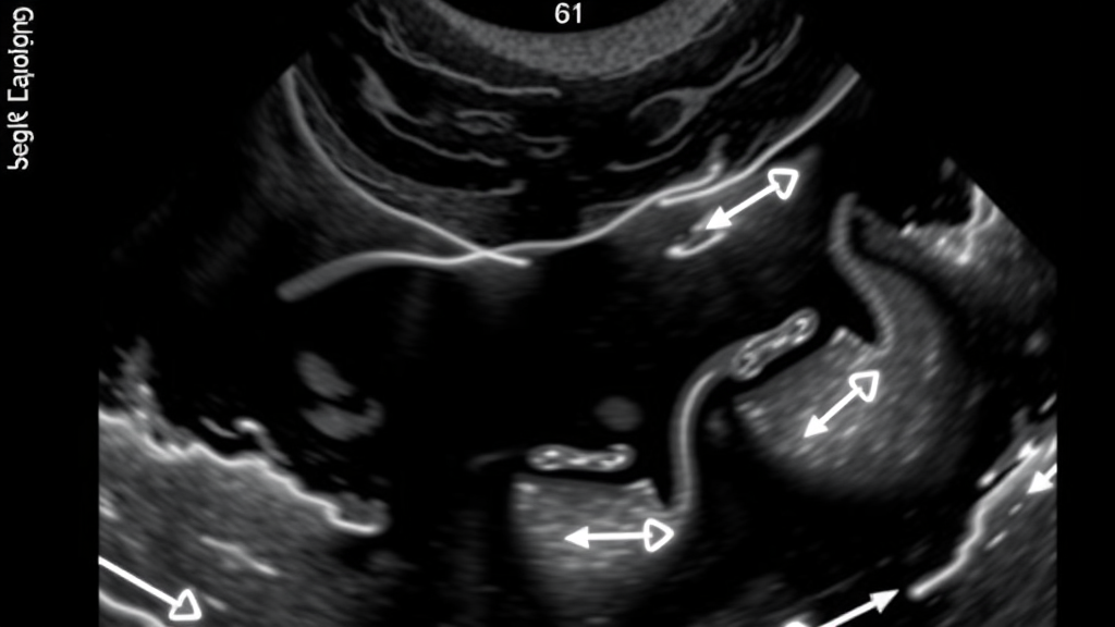
A routine kidney biopsy in a child is a procedure most parents brace for with anxiety, and the question that haunts the recovery room is deceptively simple: is the child bleeding more than expected? A new retrospective study from a Canadian pediatric hospital suggests that the answer may already be visible on the ultrasound machine before the child ever leaves the procedure suite. Researchers analyzing more than two decades of biopsy records found that a small collection of blood around the kidney, measurable immediately after the needle is withdrawn, is the single strongest signal that a child will go on to develop clinically significant hemorrhage. The finding, published in Pediatric Radiology, could reshape how clinicians monitor children after one of the most common invasive procedures in pediatric nephrology.
Percutaneous renal biopsy, in which a thin needle is guided through the skin and into the kidney to extract a sliver of tissue, is a cornerstone of modern kidney medicine. It allows pathologists to diagnose glomerular diseases, assess inflammation, gauge the severity of transplant rejection, and guide treatment decisions that can alter the trajectory of a child’s kidney health for decades. The procedure is almost always performed under real-time ultrasound guidance, which lets the operator visualize the needle path and avoid large vessels. Yet even with this precision, bleeding remains the complication that defines the procedure’s risk profile. Blood vessels in the kidney operate under high pressure, and the organ’s rich blood supply means that even a small puncture can produce a hematoma, a pocket of clotted blood that forms around the biopsy site.
Most of what medicine knows about post-biopsy bleeding comes from studies in adults. Adult nephrology literature has identified a familiar cast of risk factors: advanced age, elevated serum creatinine, high blood pressure, anemia, larger biopsy needles, and the number of passes made with the needle. Pediatric patients, however, are not simply small adults. Their kidneys are smaller, their vascular anatomy differs, their blood volume is proportionally different, and the diseases that prompt biopsy in children, from minimal change disease to transplant rejection, follow their own patterns. When the researchers behind the new study surveyed the existing literature, they found that data on bleeding risk factors in children was strikingly limited, leaving pediatric teams to extrapolate from adult evidence that may not apply.
To close that gap, a team led by Rajkumar Meena and Gali Shapira-Zaltsberg at the Children’s Hospital of Eastern Ontario in Ottawa, working with biostatisticians Nick Barrowman and Anne Tsampalieros, conducted an institutional review board-approved retrospective study covering pediatric patients who underwent native or transplant kidney biopsies between 2010 and 2024. The study spanned fourteen years of clinical practice, capturing changes in technique, needle technology, and post-procedure care protocols over time. Because the study was retrospective and used de-identified data, the ethics board waived the requirement for individual informed consent, a standard approach for research that examines existing medical records rather than prospectively enrolling patients.
The researchers defined their outcomes with unusual precision. The primary endpoints were twofold: a drop in hemoglobin of more than 15 grams per liter measured four to six hours after the biopsy, and the presence of a sizable perinephric hematoma, defined as at least five milliliters of blood collected around the kidney, as seen on an ultrasound performed immediately after the procedure. The hemoglobin threshold matters because a fall of that magnitude in a child represents a meaningful loss of circulating blood volume, one that clinicians cannot afford to overlook. The five-milliliter hematoma threshold, meanwhile, distinguishes trivial traces of blood, which are common and usually harmless, from collections large enough to signal genuine vascular injury. The association between the presence of a hematoma and a significant hemoglobin drop was assessed directly, and the team analyzed twelve preselected risk factors for their connection to these co-primary outcomes.
The study population comprised 207 children, with a median age of 11 years and an interquartile range of 7 to 14, meaning half of the patients fell between those ages. Of these, 170 children, or 82 percent, underwent biopsy of their own kidneys, while 37 children, or 18 percent, had biopsies of transplanted kidneys. Transplant biopsies are a distinct procedural scenario: the graft sits in the iliac fossa of the pelvis rather than in the flank, its vascular supply differs, and the immune context of the patient is shaped by immunosuppressive therapy. Including both biopsy types gave the researchers a broad view of real-world pediatric practice, though it also meant the analysis had to accommodate two anatomically different procedures within a single cohort.
The headline result is a strong statistical association between what the immediate post-biopsy ultrasound shows and what happens to the child’s blood count. Twenty-three patients, or 11 percent of the cohort, experienced a hemoglobin decrease of at least 15 grams per liter, with a 95 percent confidence interval of 8 to 16 percent. Twenty-seven patients, or 13 percent, developed a perinephric hematoma of at least five milliliters, with a confidence interval of 9 to 18 percent. When the researchers cross-tabulated these outcomes, they found that children with a hematoma of five milliliters or more on immediate post-procedural imaging had roughly 4.6 times the odds of experiencing a significant hemoglobin decline compared with those who did not, with a 95 percent confidence interval of 1.5 to 13.4 and a p-value of 0.004. In practical terms, a child who leaves the biopsy suite with a measurable bleed around the kidney is several times more likely to be the child whose hemoglobin falls meaningfully in the hours that follow.
Just as telling is what the study did not find. The researchers examined twelve different preselected risk factors, a list that reflects the predictors repeatedly flagged in the adult literature, and after correcting for multiple statistical testing, none of the others showed a significant association with either hemoglobin decline or hematoma formation. This negative result carries real weight. It suggests that the conventional adult risk factors, and the demographic and procedural variables clinicians might instinctively reach for, do not reliably identify which children will bleed after a pediatric renal biopsy. The immediate ultrasound image, in other words, may be more informative than any characteristic of the patient measured before the needle goes in. The finding aligns with earlier work in adult populations, including Japanese and Chinese studies that examined ultrasonographic findings and renal parenchymal thickness as predictors of post-biopsy bleeding, but it extends that logic into pediatric practice, where the evidence base had been thin.
The implications for clinical practice are concrete. If a five-milliliter hematoma on immediate post-biopsy ultrasound predicts a meaningful hemoglobin drop, then that single image could serve as a triage tool. Children with a sizable hematoma might be flagged for closer observation, repeat hemoglobin checks, longer monitoring periods, or earlier involvement of the nephrology team, while children with a clean immediate scan might be candidates for streamlined discharge pathways. This matters in pediatric settings, where minimizing time in hospital and reducing unnecessary blood draws are priorities, and where the threshold for admitting a child for overnight observation varies widely between institutions. The study also underscores the value of standardized hematoma measurement; a recent scoping review of adult practice found that reporting of perirenal hematoma size after biopsy is inconsistent, making it difficult to compare studies or pool evidence. By defining a specific volumetric threshold, the Ottawa team offers a metric that other centers can adopt and validate.
There are, of course, limits to what a single-center retrospective study can establish. The cohort of 207 patients, while respectable for a pediatric procedure study, yields relatively few bleeding events, and the wide confidence intervals around the estimates reflect that statistical uncertainty. The twelve risk factors examined were preselected, and other variables not captured in the records, such as subtle differences in technique or patient hydration status, could conceivably play a role. The hematoma-hemoglobin association, while statistically robust, is an association observed in retrospective data rather than a demonstrated causal pathway, and the finding awaits validation in independent, ideally multi-center, pediatric cohorts before it should drive wholesale changes to monitoring protocols. Still, the study’s message is clear and clinically actionable: in children undergoing ultrasound-guided kidney biopsy, the most valuable predictor of clinically significant bleeding may not be found in the chart at all, but on the ultrasound screen in the minutes immediately after the biopsy needle is withdrawn. For a procedure performed thousands of times each year in children’s hospitals around the world, that is a finding worth watching.
Subject of Research: Risk factors for clinically significant hemorrhage after ultrasound-guided percutaneous renal biopsy in children
Article Title: Predictors of clinically significant hemorrhage following ultrasound-guided percutaneous renal biopsy in pediatric patients
Article References: Meena, R., Barrowman, N., Tsampalieros, A., & Shapira-Zaltsberg, G. (2026). Predictors of clinically significant hemorrhage following ultrasound-guided percutaneous renal biopsy in pediatric patients. Pediatric Radiology. https://doi.org/10.1007/s00247-026-06794-w
Image Credits: AI Generated
DOI: 10.1007/s00247-026-06794-w
Keywords: renal biopsy, pediatric nephrology, hemorrhage, ultrasound, perinephric hematoma, hemoglobin decline, kidney transplant biopsy, interventional radiology, post-biopsy monitoring, risk factors, Pediatric Radiology, retrospective study
Cite Scienmag News
Nathaniel Bowman. (September 20, 2026). Ultrasound Right After Childhood Kidney Biopsy Predicts Dangerous Bleeding, Study Finds. Scienmag. https://scienmag.com/ultrasound-right-after-childhood-kidney-biopsy-predicts-dangerous-bleeding-study-finds/
Nathaniel Bowman. "Ultrasound Right After Childhood Kidney Biopsy Predicts Dangerous Bleeding, Study Finds." Scienmag, 20 September 2026, https://scienmag.com/ultrasound-right-after-childhood-kidney-biopsy-predicts-dangerous-bleeding-study-finds/. Accessed 20 September 2026.
Nathaniel Bowman. "Ultrasound Right After Childhood Kidney Biopsy Predicts Dangerous Bleeding, Study Finds." Scienmag. September 20, 2026. https://scienmag.com/ultrasound-right-after-childhood-kidney-biopsy-predicts-dangerous-bleeding-study-finds/


